*NURSING > STUDY GUIDE > FINAL EXAM STUDY GUIDE HEALTH CARE LATEST 2021/2022 APPROVED (All)
FINAL EXAM STUDY GUIDE HEALTH CARE LATEST 2021/2022 APPROVED
Document Content and Description Below
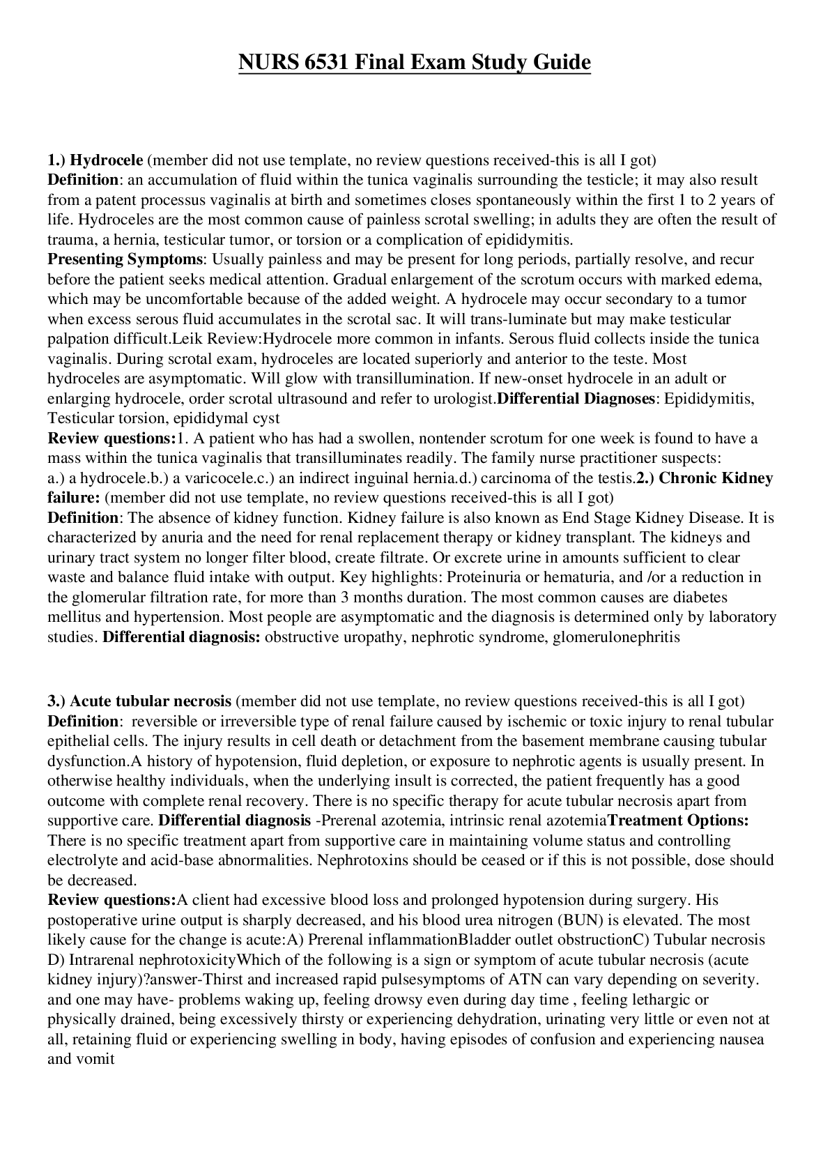
1.) Hydrocele (member did not use template, no review questions received-this is all I got) Definition: an accumulation of fluid within the tunica vaginalis surrounding the testicle; it may also res... ult from a patent processus vaginalis at birth and sometimes closes spontaneously within the first 1 to 2 years of life. Hydroceles are the most common cause of painless scrotal swelling.; in adults they are often the result of trauma, a hernia, testicular tumor, or torsion or a complication of epididymitis. Presenting Symptoms: Usually painless and may be present for long periods, partially resolve, and recur before the patient seeks medical attention. Gradual enlargement of the scrotum occurs with marked edema, which may be uncomfortable because of the added weight. A hydrocele may occur secondary to a tumor when excess serous fluid accumulates in the scrotal sac. It will transluminate but may make testicular palpation difficult. Leik Review: Hydrocele more common in infants. Serous fluid collects inside the tunica vaginalis. During scrotal exam, hydroceles are located superiorly and anterior to the testes Most hydroceles are asymptomatic. Will glow with transillumination. If new-onset hydrocele in an adult or enlarging hydrocele, order scrotal ultrasound and refer to urologist. Differential Diagnoses: Epididymitis, Testicular torsion, epididymal cyst Review questions: 1. A patient who has had a swollen, nontender scrotum for one week is found to have a mass within the tunica vaginalis that transilluminates readily. The family nurse practitioner suspects: a.) a hydrocele. b.) a varicocele. c.) an indirect inguinal hernia. d.) carcinoma of the testis. 2.) Chronic Kidney failure: (member did not use template, no review questions received-this is all I got) Definition: The absence of kidney function. Kidney failure is also known as End Stage Kidney Disease. It is characterized by anuria and the need for renal replacement therapy or kidney transplant. The kidneys and urinary tract system no longer filter blood, create filtrate. Or excrete urine in amounts sufficient to clear waste and balance fluid intake with output. Key highlights: Proteinuria or hematuria, and /or a reduction in the glomerular filtration rate, for more than 3 months duration. The most common causes are diabetes mellitus and hypertension. Most people are asymptomatic and the diagnosis is determined only by laboratory studies. Differential diagnosis: obstructive uropathy, nephrotic syndrome, glomerulonephritis 3.) Acute tubular necrosis (member did not use template, no review questions received-this is all I got) Definition: reversible or irreversible type of renal failure caused by ischemic or toxic injury to renal tubular epithelial cells. The injury results in cell death or detachment from the basement membrane causing tubular dysfunction. A history of hypotension, fluid depletion, or exposure to nephrotic agents is usually present. In otherwise healthy individuals, when the underlying insult is corrected, the patient frequently has a good outcome with complete renal recovery. There is no specific therapy for acute tubular necrosis apart from supportive care. Differential diagnosis -Prerenal azotemia, intrinsic renal azotemia Treatment Options: There is no specific treatment apart from supportive care in maintaining volume status and controlling electrolyte and acid-base abnormalities. Nephrotoxins should be ceased or if this is not possible, dose should be decreased. Review questions: A client had excessive blood loss and prolonged hypotension during surgery. His postoperative urine output is sharply decreased, and his blood urea nitrogen (BUN) is elevated. The most likely cause for the change is acute: A) Prerenal inflammation Bladder outlet obstruction C) Tubular necrosis D) Intrarenal nephrotoxicity Which of the following is a sign or symptom of acute tubular necrosis (acute kidney injury)? answer-Thirst and increased rapid pulse symptoms of ATN can vary depending on severity. and one may have- problems waking up, feeling drowsy even during day time , feeling lethargic or physically drained, being excessively thirsty or experiencing dehydration, urinating very little or even not at all, retaining fluid or experiencing swelling in body, having episodes of confusion and experiencing nausea and vomit 4. Indirect inguinal hernia Definition: Indirect inguinal hernia – Indirect inguinal hernia is caused by a birth defect in the abdominal wall that is present at birth. A scrotal-inguinal hernia results when a segment of the bowel slips through the internal inguinal ring, where it may remain in the inguinal canal or pass into the scrotal sac. An inguinal hernia may occur as a result of a defect in the anterior abdominal wall or because of a patent process vaginalis. Inguinal hernias predominantly affect men (9:1) and have the highest incidence in men aged 40 to 59. A hernia may move freely between the abdomen and the scrotum or can be spontaneously reduced by digital manipulation. When a hernia becomes strangulated or is unreducible, this compromises the blood supply and requires emergent surgical reduction. Strangulation should be suspected when a tender mass is palpated in the scrotum in addition to redness, nausea, and vomiting Presenting Symptoms: Scrotal swelling, mild to moderate pain on straining, scrotal heaviness, and the possible presence of a bulge are common complaints. Increased edema after standing in an erect position but decreases when the patient is recumbent. 3 Differential Diagnoses: undescended testis, lymphadenopathy, femoral hernia Pattern Recognition: Enlarged hemiscrotum or a bulge in the groin area that may spontaneously reduce when the patient is supine or with manual reduction. The provider will not be able to move the fingers above the mass, which should be soft and mushy but painless unless it is incarcerated and ischemic. Scrotal hernias do not transilluminate. Auscultation of bowel sounds over the mass is significant for the diagnosis of bowel in the scrotal sac. Treatment options: If the herniated bowel is reducible, surgical referral for possible future repair is indicated. Difficulty in reducing a hernia is cause for urgent surgical intervention. However, pain may indicate incarceration of the bowel or complete inability to reduce the hernia, which is cause for immediate emergency department referral and surgical exploration. Review questions: 1. Mr. S. comes to you with scrotal pain. The examinations of his scrotum, penis, and rectum are normal. Which of the following conditions outside of the scrotum may present as scrotal pain? A. Inguinal herniation and peritonitis ** B. Renal colic and cardiac ischemia C. Pancreatitis and Crohn ’ s disease D. Polyarteritis nodosa and ulcerative colitis Rationale: Conditions outside of the scrotum that may present with scrotal pain are abdominal aortic aneurysm, inguinal herniation, pancreatitis, renal colic, peritonitis, intraperitoneal hemorrhage, and polyarteritis nodosa. Keep in mind that any client with scrotal pain should be considered to have testicular torsion until proved otherwise, especially in the age groups of the neonate and adolescents. 2. The most common type of hernia is a(n): A. indirect inguinal hernia. ** B. direct inguinal hernia. C. femoral hernia. D. umbilical hernia. Rationale: An indirect inguinal hernia is the most common type of hernia affecting all ages and both genders and accounts for 50% of hernias treated. The point of origin is above the inguinal ligament and often travels into the scrotum. A direct inguinal hernia is less common (accounts for about 25% of hernias seen) and usually occurs in men older than age 40. The point of origin is above the inguinal ligament and rarely travels into the scrotum. The femoral hernia is the least common (about 10% of hernias seen) and occurs more often in women than in men. The point of origin is below the inguinal ligament and never travels into the scrotum in men. An umbilical hernia occurs more frequently in infants and is a protrusion of part of the intestine at the umbilicus. 3. Max, age 70, is obese. He is complaining of a bulge in his groin that has been there for months. He states that it is not painful, but it is annoying. You note that the origin of swelling is above the inguinal ligament directly behind and through the external ring. You diagnose this as a(n): A. indirect inguinal hernia. B. direct inguinal hernia. ** C. femoral hernia. D. strangulated hernia. Rationale: A direct inguinal hernia usually occurs in middle-aged to older men and is the result of an acquired weakness caused by heavy lifting, obesity, or chronic obstructive pulmonary disease (COPD). The origin of swelling is above the inguinal ligament directly behind and through the external ring. An indirect inguinal hernia is congenital or acquired and is more common in infants younger than 1 year of age and in men ages 16 – 25. The origin of swelling is above the inguinal ligament. The hernia sac enters the canal at the internal ring and exits at the external ring. A femoral hernia, which occurs more frequently in women, is acquired and results from an increase in abdominal pressure, as well as muscle weakness. The origin of swelling is below the inguinal ligament. Because Max is not having any pain and the condition has been this way for months, you know that the hernia is not strangulated. A strangulated hernia, which requires immediate referral to a surgeon, results in no blood supply to the affected bowel and causes nausea, vomiting, and tenderness. 5. Orchitis Definition: Orchitis is a systemic, blood-borne infection that results in an acute inflammation of one or both testicles. It may coexist with infections of the prostate and epididymis; causes – viral infection (ex. Mumps), C. trachomatis and N. gonorrhoeae in adolescents, E. coli – men, complication of syphilis, mycobacterial, fungal; hydrocele and scrotal wall thickening may be seen as a complication of mumps Presenting Symptoms: Gradual onset of acute or moderate pain, testicular swelling, and fever 3 Differential Diagnoses: epididymitis, testicular tumor, hernia, testicular torsion Pattern Recognition: Testicular edema may be so pronounced that it is difficult to distinguish the testes from the epididymis. Palpation may reveal swollen, very tense testes that are painful, and the patient may be febrile. Inflammation of the testis usually involves systemic viral infections (commonly mumps) and includes unilateral or bilateral erythema, edema, and scrotal tenderness, which occurs 4 to 7 days after initial fever. Treatment options: Anti-infective therapy is recommended, with guidance by local sensitivity reports. The following antibiotic regimens are effective against the most common causes of epididymitis: singledose ceftriaxone given intramuscularly (IM), 250 to 500 mg, and doxycycline, 100 mg twice daily for 10 days for men younger than 35 years; in men older than 35 years, levofloxacin (given intravenously [IV] or orally [PO]), 500 to 750 mg/day, or ciprofloxacin, 500 mg (IV or PO), for 10 to 14 days. Antipyretics should be used to reduce discomfort and fever, and an anti-inflammatory agent should be prescribed. An antiemetic can also be prescribed for nausea and vomiting. Bed rest and scrotal elevation are also recommended for epididymitis. Hot or cold compresses may be helpful for orchitis. Review questions: A 35 year old sexually active man presents with a 1 week history of fever and pain over the left scrotum. It is accompanied by frequency and dysuria. The scrotum is edematous and tender to touch. He denies flank pain, nausea, and vomiting. He reports that eh pain is lessend when he uses scrotal-support briefs. The urinalysis shows 2 + blood and a large number of leukocytes. What is the most likely diagnosis? A. Acute urinary tract infection B. Acute pyelonephritis C. Acute orthitis D. Acute epididymitis ** Orchitis is caused by which of the following? A. Mumps virus ** B. Measles virus C. Chlamydia trachomatis D. Chronic urinary tract infections that are not treated adequately A 10 year old boy complains of sudden onset of scrotal pain upon awakening that morning. He is also complaining of severe nausea and vomiting. During the physical examination, the nurse practitioner finds a tender, warm, and swollen left scrotum. The cremastic reflex is negative and the urine dipstick is negative for leukocytes, nitrites, and blood. The most likely diagnosis is: A. Acute epididymitis B. Severe salmonella infection C. Testicular torsion ** D. Acute orchitis What type of follow up should this patient receive? A. Refer to a urologist within 48 hours B. Refer him to the emergency department as soon as possible ** C. Prescribe ibuprofen (advil) 600 mg QID for pain D. Order a testicular ultrasound for further evaluation 6. Testicular torsion Definition: Testicular torsion - obstruction of blood flow to the testes because of a twisting of the arteries and veins in the spermatic cord resulting in occlusion of blood flow. Occurs in 12-18 year olds. Usually unilateral, effecting the left testis. Two types: extravaginal and intravaginal. Extravaginal (rare, seen in neonates)- twisting of the spermatic cord, testis, and process vaginalis; intravaginal (seen in adolescents)- failure of the testis to adhere to the scrotal wall, creating a “bell clapper deformity.” Different from torsion of the appendix testis. Presenting Symptoms: sudden in onset, extremely painful, and may awaken the patient from sleep or be trauma induced. Testicular pain, experience abdominal pain, nausea, and vomiting; 25% of patients have a fever. Clinical manifestations-testicle that rides high in the scrotum and an absent cremasteric reflex on examination 3 Differential Diagnoses: testicular appendix torsion, epididymitis, epididymo-orchitis, hydrocele Pattern Recognition: Most common in the left hemiscrotum. Scrotal edema and erythema may be seen. The affected side may have a higher position as a result of rotation. The spermatic cord is swollen and extremely tender, the epididymis may be felt anteriorly, and the majority of patients will have an absent cremasteric reflex. In some instances a small area of cyanosis (blue dot sign) may be present on the scrotal skin and indicates torsion of the appendix testis. Treatment options: Surgical consultation with surgical exploration – needs to occur in 6 hours. Review questions: 1. A 24-year-old man presents with sudden onset of left-sided scrotal pain. He reports having intermittent unilateral testicular pain in the past but not as severe as this current episode. Confirmation of testicular torsion would include all of the following findings except: A.unilateral loss of the cremasteric reflex. B.the affected testicle held higher in the scrotum. C.testicular swelling. D.relief of pain with scrotal elevation. ** [Show More]
Last updated: 1 year ago
Preview 1 out of 92 pages
Reviews( 0 )
Document information
Connected school, study & course
About the document
Uploaded On
May 14, 2022
Number of pages
92
Written in

Additional information
This document has been written for:
Uploaded
May 14, 2022
Downloads
0
Views
38


.png)
.png)


.png)