*NURSING > STUDY GUIDE > Patho NR283-Exam 1 Study Guide,100% CORRECT (All)
Patho NR283-Exam 1 Study Guide,100% CORRECT
Document Content and Description Below
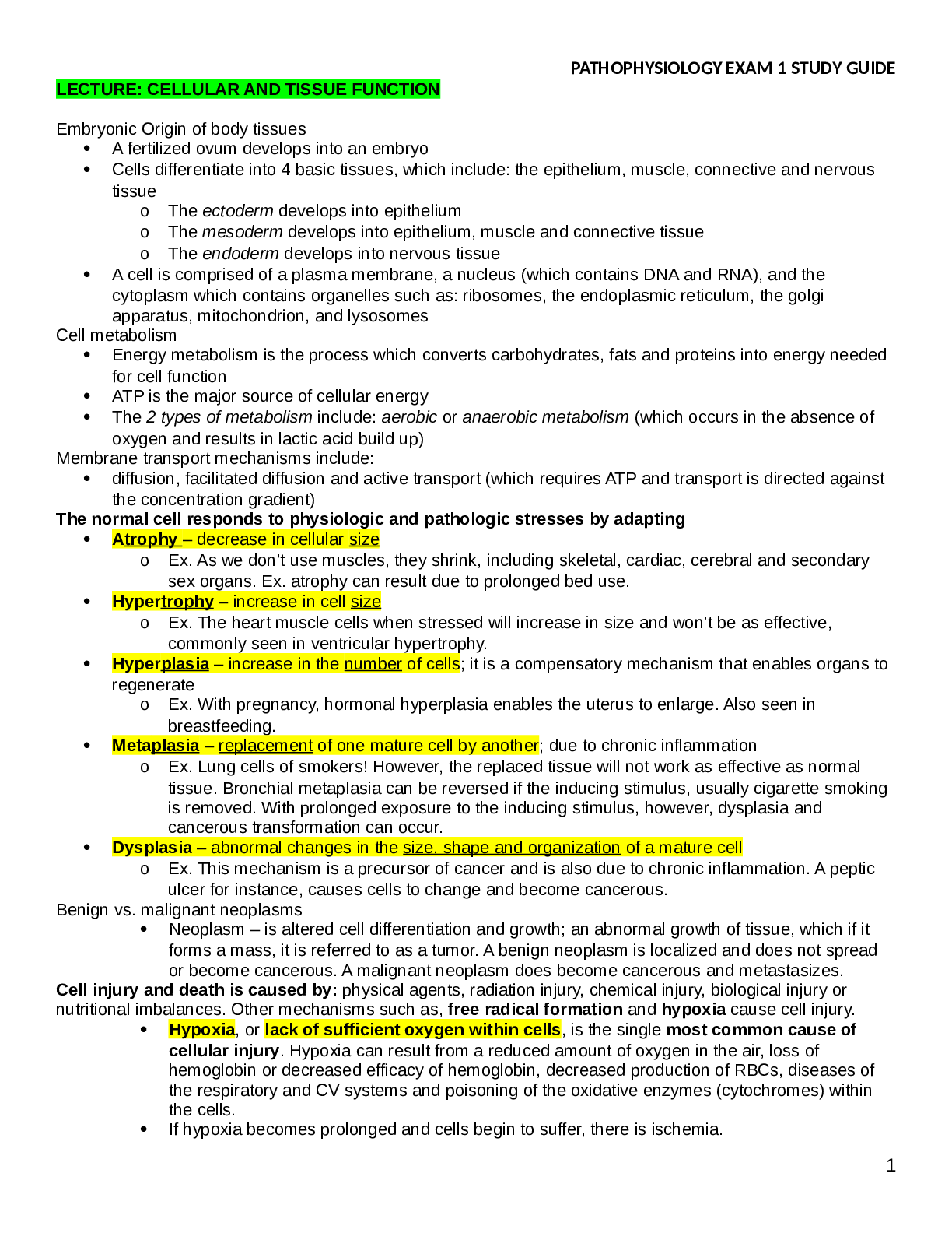
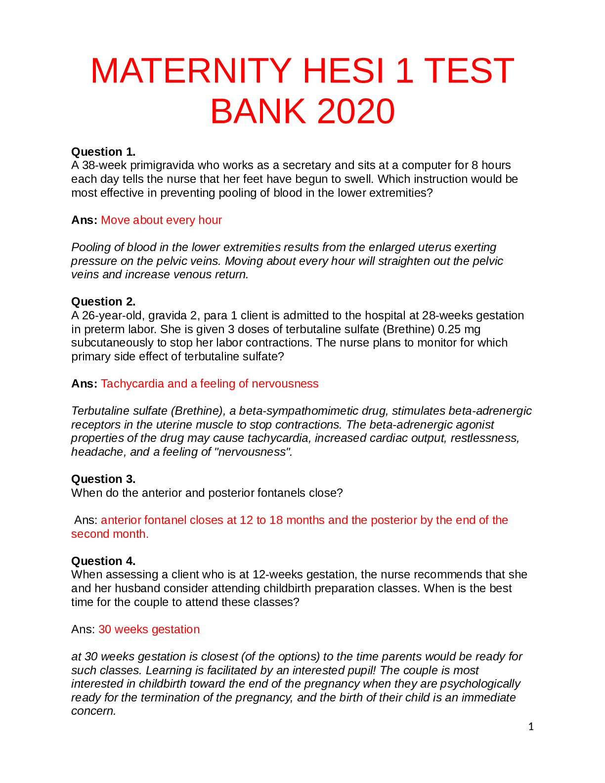
Patho NR283-Exam 1 Study Guide Chapter 1 Atrophy: ▪ Decrease in the size of cells ▪ Results in reduced tissue mass ▪ Common causes: reduced use of the tissue (in cast), insufficient nutri... tion, decrease neurologic or hormonal stimulation, aging Hypertrophy: ▪ Increase in cell size ▪ Results in enlarged tissue mass; ex) enlarged heart w/ heart failure ▪ Causes: additional work by the tissue( lifting weights), excessive hormonal stimulation Hyperplasia: ▪ Increased number of cells ▪ Results in enlarged tissue mass ex) uterus in pregnancy ▪ Causes: compensatory mechanism to meet increased demand, or pathologic when there is a hormonal imbalance Metaplasia: ▪ Mature cell type is replaced by a different mature cell type ▪ Adaptive mechanism that provided more resistance tissue Dysplasia: ▪ Cells vary in size and shape within a tissue ▪ Chronic irritation infection, or it may be a precancerous change Anaplasia: ▪ Undifferentiated cells with variable nuclear and cell structures ▪ Characteristic of cancer Neoplasia: ▪ New growth ▪ Commonly called tumor Causes of cell death: 1. Ischemia: Most common cause of cell death; Decreased supply of oxygenated blood to a tissue or organ due to circulation obstruction 2. Hypoxia: Reduced oxygen in tissues/blood; Result insufficient oxygen and reduced cellular metabolism 3. Physical damage: extreme heat/ cold 4. Mechanical damage: pressure, tumor, obstruction 5. Chemical toxins: exogenous-pollution endogenous-free radicals 6. Microorganisms/pathogens -bacteria/viruses Stages 1. Initial: start; loss of function; reversible 2. Irreversible: Cell death Aptosis: ▪ Normal; refers to programmed cell death Necrosis: ▪ Group of cells die due to cell injury Gangrene: ▪ Area of necrotic tissue that has been invaded by bacteria Chapter 20 Differentiation: ▪ Each cell type differentiates/matures and carries out specific functions ▪ The structure reflects the function of the tissue Benign: ▪ Have tissue name plus the suffix –oma ▪ Differentiated cells that reproduce at a higher rate but still normal; slower growth; ▪ Do NOT spread ▪ Localized ▪ Encapsulated ▪ Tissue damage to adjacent cells from pressure ▪ Rare systemic manifestations Malignant: ▪ Have the tissue name plus the suffix –carcinoma ▪ Sarcomas: tumor of the connective tissue... Often malignant ▪ Undifferentiated, nonfunctional cells ▪ Rapid reproduction ▪ Infiltrate and spread to surrounding tissue ▪ No Capsule ▪ Spread to different sites ▪ Normal cell destruction ▪ Systemic manifestation present Cancer risk factors: 1. Environment 2. Genetics 3. Infections 4. Chemicals/toxins 5. Biological factors 6. Age 7. Diet 8. Hormones (^ estrogen levels= breast cancer) 1. Unusual bleeding or discharge anywhere in the body 2. Change in bowel or bladder habits 3. Change in wart or mole 4. A sore that does not heal 5. Unexplained weight loss 6. Anemia, low hemoglobin and persistent fatigue 7. Persistent cough or hoarseness without reason 8. A solid lump; in the breast/testes, or anywhere in the body Local Effects of Tumors: ▪ Pain: o May be absent until very late stages o Occurs when tumor is well advanced o Severity depends on the type of tumor ▪ Obstruction: o Occurs when tumor compresses a duct or passageway o Blood supply or lymphatic flow may be restricted o Digestive tract o Airflow in bronchi ▪ Tissue Necrosis/Ulceration: o May lead to bleeding or infection around the tumor (ischemia necrosis) Systemic Effects of Malignant Tumors: ▪ Weight loss and (weakness and wasting): o Anorexia, fatigue, pain, stress o Increased demands on the body from tumor cells ▪ Anemia: o Caused by blood loss at tumor site o Nutritional deficits may reduce hemoglobin synthesis ▪ Severe fatigue: o Caused by inflammatory changes, cachexia and anemia o Stress of treatment schedule o Psychological factors ▪ Infections: o Often occur as resistance declines ▪ Bleeding: o Tumor cells may erode the blood vessels/platelets ▪ Paraneoplastic syndrome: o Associated with certain tumor types o Tumor cells release substances that affect neurological function and may have hormonal effects Spread: 1. Invasion: • Local spread • Tumor cells grow into adjacent tissues • Example: uterine carcinoma invades the vagina 2. Metastasis: • Spread to distant sites • Via blood or lymph or other body fluids 3. Seeding: • Spread of cancer cells in body fluids or along membrane; usually in body cavities • Example: carcinoma of the colon spreads to the liver Staging: ▪ Describes the extent of the disease ▪ Used to estimate prognosis ▪ TMN system o Size of primary tumor (T) o Involvement of regional lymph nodes (N) o Spread of tumor (M)-metasistes ▪ Stage 1: small, localized, good prognosis ▪ Stage IV: well advanced, difficult to treat, spread, poor prognosis Diagnostic tests: • Blood test • Tumor markers • Imaging • Cytologic tests (for screening high risk individuals); Confirms cancer via biopsy Treatment • Chemo • Radiation • Gene therapy • Stem cell transplant • Drugs Chapter 2 Filtration ▪ Movement into interstitial (filtering goods from blood into the space for the cell) Osmosis ▪ Movement into intervascular Fluid Volume Excess Causes 1. Kidney failure 2. Liver failure 3. Heart failure 4. Increased sodium intake 5. IV fluids 6. Blood transfusions Manifestations 1. Dyspnea 2. Decreased lab values (everything is diluted) 3. Increased urine output 4. High BP 5. JVD 6. Edema Edema: • Excessive amount of fluid in the interstitial compartment, which causes a swelling or enlargement of the tissue Causes of Edema: 1. Increased capillary hydrostatic pressure o Caused by higher bp and increased blood volume o Forces increased fluid out of capillaries into tissue o Cause of pulmonary edema 2. Decreased plasma osmotic pressure o Results in loss of plasma proteins- particularly albumin o Risks: liver failure; burns pt; malnutrition 3. Obstruction of lymphatic circulation o Causes localized edema o Excessive fluid and protein not returned to general circulation 4. Increased capillary permeability o Usually causes localized edema o May result in an inflammatory response or infection o Histamines and other chemical mediators increase capillary permeability o Can also result from bacterial toxins or large burn wounds and result in widespread edema Complications of Edema: • Swelling o Red or pale in color • Pitting edema o Depression remains when finger pressure is removed 1. Functional impairment -Restricts range of joint movement 2. Pain 3. Decreased arterial circulation 4. Ischemia leading to tissue breakdown, tissue necrosis and ulcers -Edematous tissue in skin is susceptible; Proper skin care Examples: • Exposure to allergens ▪ Increased capillary permeability and release of histamines • Arm swelling after mastectomy ▪ Lymphatic obstruction • Lack of albumin causes abdominal swelling ▪ Decreased plasma osmotic pressure • Swelling of ankles associated with heart problems ▪ Increased hydrostatic pressure Dehydration: ▪ Insufficient body fluid resulting from inadequate intake or excessive loss Causes of Dehydration: ▪ Vomiting and diarrhea ▪ Excessive sweating with loss of sodium and water ▪ Insufficient water intake in older adults or unconscious persons Effects of Dehydration: ▪ Dry mucous membranes in the mouth ▪ Decreased skin turgor or elasticity ▪ Lower blood pressure ▪ Tachycardia ▪ Decreased mental function or confusion ➔ Review of Sodium ◆ Normal lab value: 135-145mEq/L ◆ The main electrolyte in the extracellular fluid (ECF) ◆ Primarily controlled by the kidneys via Aldosterone ◆ Sodium Linked with Neuro (Cerebral edema) ◆ Essential for ● Nerve impulses ● Muscle contraction ● Osmotic pressure ◆ Sources of sodium- food and beverage ◆ Excreted through sweat, urine, and feces Sodium Imbalance ➔ Review of Potassium ◆ Normal lab values: 3.5-5mEq/L ◆ The main electrolyte in intracellular fluid ◆ Linked with the heart ◆ Essential for ● Nerve impulses ● Muscle contractions ◆ Sources of potassium: food and beverage ◆ Excreted through urine and feces Potassium Imbalance Hypokalemia <3.5 Hyperkalemia >5.0 Causes 1. Diuretics 2. Inadequate K+ intake 3. Excessive diarrhea Manifestations 1. Muscle fatigue/cramps 2. nausea/vomiting/ 3. Constipation 4. Cardiac dysrhythmias Causes 1. Renal failure 2. K+ sparing diuretics 3. Burns/crush injuries Manifestations 1. Muscle weakness( paralysis) 2. Paresthesias 3. Cardiac dysrhythmias ( cardiac arrest) ➔ Review of Calcium ◆ Normal lab values: 8.5-10.5mEq/L ◆ Linked with bones and muscle ◆ Stored in bones and remaining in extracellular fluid ◆ Controlled by the parathyroid hormone (PTH) and Calcitonin ◆ Vitamin D promotes the movement of Ca+ from bone and intestines to the blood ◆ Essentials for ● Muscle contractions ● Strength of teeth and bones ● Stability of nerve membranes ◆ Sources of calcium include milk and milk products ◆ Excreted through urine and feces Calcium Imbalances Hypocalcemia <8.5 Hypercalcemia >10.5 Causes 1. Hypoparathyroidism 2. Malabsorption 3. Vitamin D deficiency Causes 1. Hyperparathyroidism 2. Bone cancer 3. Immobility Manifestations 1. Muscle twitching 2. Tetany 3. Paresthesias 4. Chvostek and trousseau signs 5. Cardiac dysrhythmias Manifestations 1. Muscle weakness 2. Loss of muscle tone 3. Spontaneous fractures 4. Kidney stones (Renal Calculi) 5. Cardiac dysrhythmias ➔ Review of Magnesium ◆ Normal lab value: 1.6-2.5mg/dL ◆ 50% located in the intracellular fluid, 50% located in the bone ◆ Linked with CNS ◆ Essential for ● Protein and DNA synthesis ● ATP production ● Nerve conduction ● Relaxes smooth muscle ◆ Independent relationships with K+, Ca+ ● If magnesium is low K+ and Ca+ are low ◆ Sources of magnesium: food ◆ Excreted through urine Magnesium imbalance Hypomagnesemia <1.6 Hypermagnesemia >2.5 Causes 1. Malnutrition issues 2. Malabsorption issues 3. Diuretics Manifestations 1. Tremors 2. Hyperreflexia (can’t relax so its tense) 3. Insomnia Causes 1. Renal failure 2. Increased magnesium intake Manifestations 1. Hyporeflexia 2. Lethargy 3. Respiratory depression Acid-Base: ▪ Normal pH range 7.35-7.45 ▪ Co2: 35-45 ▪ HCO3: 22-26 ▪ Acidosis o Excess hydrogen ions/CO2/water, decrease in pH ▪ Alkalosis o Deficit of hydrogen ions, increase in pH ▪ Respiratory system can alter carbonic acid levels to change pH ▪ Kidneys can modify the excretion rate of acids and absorption of bicarbonate to regulate pH o Most significant control mechanism o Slowest mechanism Respiratory Acidosis Respiratory Alkalosis Causes -Hypercapnia -Hypoventilation Risk 1. -Opiate overuse/overdose 2. -Respiratory disease 3. -Sleep apnea 4. -Airway obstruction 5. -Anesthetics Manifestation (^ CO2 creates vasodilation- brain depression) 1. -Headache 2. -Confusion 3. -Lethargy 4. -Coma 5. -Tremors 6. -Paralysis Causes -Hypocapnia -Hyperventilation Risk 1. Panic attacks 2. Fever 3. Brain injuries 4. Mechanical ventilation Manifestations (Vasoconstriction- brain is flustered/overactive) 1. Agitated 2. Irritable 3. Light-headed 4. Tetany 5. Paresthesis 6. Seizures Metabolic Acidosis Metabolic Alkalosis Causes - Gain in H+ (Acid) - Loss of HCO3 Risk Gain in acid: 1. Salicylate toxictity 2. Diabetic ketoacidosis 3. Renal failure Causes - Loss of H+ - Gain in HCO3 Risk Loss of acid: 1. Vomiting 2. GI tube suctioning 3. Diuretics 4. Strenuous exercise (lactic acid) 5. Sepsis Decrease in HCO3: 6. Diarrhea Manifestations 1. Headache 2. Confusion 3. Lethargy 4. Coma 5. Kussmaul’s respirations 6. Nausea/vomiting/abdominal pain 7. Cardiac dysrhythmias Gain in HCO3 4. Excess intake of antacids Manifestations 1. Agitation 2. Irritable 3. Light-headedness 4. Tetany 5. Paresthesias 6. Seizures Chapter 5 First Line of Defense: ▪ Nonspecific ▪ Mechanical barrier o Unbroken skin and mucous membranes o Secretions such as tears and gastric juices Second Line of Defense: ▪ Nonspecific ▪ Phagocytosis ▪ Inflammation Third Line of Defense: ▪ Specific disease ▪ Production of specific antibodies and cell-mediated immunity Inflammation: ▪ A normal protective mechanism (second line of defense) ▪ Localizes and removes injurious agents ▪ Disorders ending in –itis ▪ Not always a sign of infection o Infection is one cause of inflammation ▪ Signs and symptoms serve as warning sign o Problem may be hidden within the body Causes of Inflammation: 1. Direct physical damage 2. Caustic chemicals 3. Ischemia or infarction 4. Allergic reactions 5. Extreme hot or cold 6. Foreign bodies 7. Infection Steps of Inflammation: ▪ 1. Injury to capillaries and tissue cells ▪ 2. Release of chemical mediators (histamine, prostaglandins, bradykinin) ▪ bradykinin from injured cells ▪ bradykinin stimulates pain receptors ▪ Pain causes release of histamine ▪ Bradykinin and histamine cause 3. capillary dilation ▪ 4. Capillary permeability ▪ 5. Leukocytes move to injury site- Migration of neutrophils and monocyte to the site of injury ▪ 6. Phagocytosis occurs. Neutrophils phagocytize bacteria; Macrophages leave the bloodstream and phagocytose microbes Local Effects of Inflammation: ▪ Redness and ▪ Warmth o Caused by increased blood flow to the damaged area ▪ Swelling o Shift of protein and fluid into interstitial space ▪ Pain o Increased pressure of fluid on nerves o Release of chemical mediators (bradykinins) ▪ Exudate o Collection of interstitial fluid formed in the inflamed area Systemic Effects of Inflammation: ▪ Mild fever ▪ Malaise ▪ Fatigue ▪ Anorexia Acute Inflammation: ▪ Process of inflammation is the same regardless of cause ▪ Timing varies with specific cause ▪ Vasodilation ▪ Hyperemia o Increased blood flow in the area ▪ Increase in capillary permeability o Plasma proteins move into interstitial space ▪ Chemotaxis to attract cells of the immune system Chronic Inflammation: Causes: • When causative factors of acute are not removed • Chronic irritation -Smoking -long-term immune response Complications ▪ Deep ulcers may result ▪ Perforations ▪ Cell necrosis and lack of regeneration ▪ Periodic exacerbations of acute inflammation Lab Tests • CBC-WBC, Leukocytes • CRP (c-reactive protein) • ESR or Sed rate Burn Classification: DAPH ▪ Depth of skin damage o First degree: -superficial burns(outer layer of epidermis) -Red/pink -Very painful -no skin damage 3-10 repair o Second degree: Superficial-Partial: -involve damage to all epidermis and part of dermis -red/blistered/moist -very painful /sensitive -1-2 weeks complete repair Deep Partial: All epidermis & dermis -mottled/pink-redish/waxy white -numbness/tingling – 1 month o Third degree: Full-thickness -down to subcutaneous, muscle, fascia or bone- charred/black,deep red/leathery -no pain -skin graft ▪ Percentage of body surface o Rule of nines-measures extend of burns/ allows to calculate fluid replacement Complication of Burns: ▪ Hypovolemic shock ▪ Respiratory problems ▪ Pain ▪ Infection ▪ Increased metabolic needs for healing process Healing of Burns: ▪ Hypermetabolism ▪ Immediate covering of a clean wound to prevent infection ▪ Healing is a prolonged process ▪ Scar tissue develops; even with skin grafting ▪ Physiotherapy and occupational therapy ▪ Surgery may be necessary to release scar tissue Types of Healing 1. Resolution: minimal tissue damage (ex: paper cut) 2.Regeneration: proliferation& damage (ex: surgical incision) 3. Replacement : tissue damage cells – wound is replaced w/ scar tissue Process of Healing: 1. Hemostasis -1 hourish - control of blood loss 2. Inflammatory phase 4-6 days 3. Proliferative Phase- 3-24 days filling the space 4. Maturation/remodeling 3 weeks after injury – up to 1 year – regeneration or replaced w/ scar tissue Chapter 6 Terminology ▪ Opportunistic pathogens- capable of infection in ppl w/ bad health ▪ Virulence- disease producing microbes ▪ Infectious disease result of invasion of microbes that results in manifestations Types of microorganisms 1. Bacteria -does Not require living host -Gram – or + to detect -grouped based on shape 2. Viruses -Requires living host- uses cells DNA to replicate 3. Fungi -usually don’t cause infection unless pt. is immunocompromised 4. Protoza -Parasites living independently 5. Helminths -worms that can cause infection Chain of Infection 1. Agent: The microbe causing the infection 2. Reservoir: - Environmental source such as contaminated soil - Infected person or animal 3. Portal of Exit: -Where the agent leaves the reservoir 4. Mode of Transmission: -Method/way agent reaches a new susceptible host - Air, water, direct contact, food 5. Portal of Entry: -Access to a new host 6. Susceptible Host: -Health status, immunity, age, nutrition Modes: Direct Contact: • Touching infectious lesion, sexual activity • Contact with infected blood or bodily secretion Indirect Contact: • Contaminated hand or food • Fomite o Inanimate object Droplet Transmission: • Respiratory or salivary secretions are expelled from infected individual Aerosol Transmission: • Involve small particles from the respiratory tract o Suspended in air and can travel farther than droplets o Example: TB Vector-borne • Insect or animal is an intermediate host Nosocomial: infection that occurs in healthcare facility ex: pneumonia, UTI, MRSA, surgery Standard/Universal Precautions: • Assume everyone is infected • Used in all settings with all patients when body fluids may be exchanged Specific Precautions: • In patients diagnosed with a particular infection o Used in addition to standard precautions Incubated Period: • Time between entry of organism into the body and appearance of clinical signs of disease • Vary considerable with different organisms Prodromal Period: “being DRAMATIC” • Fatigue, loss of appetite, headache • Nonspecific o “coming down with something” • More evident in some infections than others Acute Period: • Infectious disease develops fully Recovery Period: Manifestation subside- body recovers Patterns of Infection: • Local infections • Focal infections: local other tissue • Systemic infections • Mixed infections: several diff microbes at 1 site • Acute infection • Chronic infections • Primary infections: initial microbe exposure • Secondary infections: opportunist microbe causes infection • Superinfections: microbes resisted to drug therapy Manifestations of infection: Local: • Redness • Warmth • Swelling • Pain • Lymphadenopathy • Exudate Systemic: • Fever • Fatigue • Weakness • Anorexia • Leukocytosis Methods of Diagnosis: • Culture and staining • Blood tests o Leukocytosis: bacterial infection o Leukopenia: viral infection • Differential count o Neutrophil increase with acute infection o Lymphocyte and monocyte increase with chronic infection • C-reactive protein • Erythrocyte sedimentation rate • Radiologic tests Chapter 7 Antigens: • Foreign substance • Microbe or component of the cell that stimulates immune response Antibody: • Specific protein produced in humoral response to bind with antigen Autoantibody: • Antibodies against self-antigen • Attacks bodys own tissues Macrophages: • Initiation of the immune response • Engulf foreign material/ phagocytosis Lymphocytes: • One of leukocyte from bone marrow o T lymphocyte: cell mediated immunity ▪ WBC o B lymphocyte: humoral immunity ▪ Antibody producing plasma cell or a B memory cell • Natural killer cell o Destroy foreign cells, virus-infected cells and cancer cells Chemical mediators: -histamines, bradykins, prostaglandins stimulate inflammation, vasodilation IgG: • Most common antibody • Both primary and secondary immune responses • Mom to baby placenta • Hypersensitivity type 1 IgM: • First to increase in immune response • Type 2 hypersensitivity IgA: • In secretions; breastmilk IgE: • Allergic response • Bind to mast cells in skin and mucous membranes • Release of histamine and other chemicals • Results in inflammation IgD: • Attached to B cells • Activates B cells Process of Acquiring Immunity Steps of the immune response ▪ Primary ▪ First exposure to an antigen or microbe or anything that attacks the body ▪ Builds antibodies o Secondary ▪ Repeat exposure to the same antigen, the response is faster because the body already recognizes the microbe ▪ Rapid response Types of Immunity • Active- think of antigen o Active artificial ▪ immunization o Active natural ▪ When someone is exposed to the antigen and they get sick • Passive- think of antibodies o Passive artificial ▪ Inject antibodies to boost immunity when immunity is low; IVIG o Passive natural ▪ Mom and baby, when the antibodies pass through the placenta or the breast milk (IgM) Tissue and Organ Transplant Rejection o Replacement of damaged tissues or organs by healthy donors o Skin, bones, corneas, organs, bone marrow o Different types ▪ Human to human ▪ Animal to human ▪ Identical twin to twin ▪ Skin graft- one part of the body to another area • Rejection ▪ Type IV cell-mediated hypersensitivity reaction ▪ T lymphocytes see it as foreign • Types of rejection ▪ Hyperacute- immediate ▪ Acute- up to several weeks after ▪ Chronic- up to months after • Treatment ▪ Immunosuppression therapies to reduce immune response and prevent rejection ▪ Medications • Hypersensitivity Reactions (ACID) o Type I: Allergic Reactions ▪ Exposure to an allergen ▪ IgE mediated ▪ Manifestations ▪ Itching ▪ Swelling ▪ Sneezing ▪ Hives o Complications ▪ Anaphylaxis shock ▪ Airway swelling • Type II: Cytotoxic Hypersensitivity o Reaction to a cell surface antigen o Example: Blood transfusion/Wrong blood type ▪ IgG or IgM mediated ▪ Activation of the complement system o Results in ▪ Phagocytosis ▪ Lysis and destruction of the cell o Manifestations ▪ Depends on the disorder it causes • Type III: Immune Complex o Example: Lupus o Antigen and antibody combine ▪ Forms an immune complex o Immune complex ▪ Activates complement system ▪ Inflammation • Deposits in tissue, blood vessels walls ▪ Tissue and blood vessel destruction • Manifestations ▪ Depends on the disorder it causes • Type IV: Cells Mediated or Delayed o Example: TB Test ; Organ transplant o Delayed response • Sensitized T lymphocytes reaction to an antigen • Results in ▪ Release of lymphokines ▪ Inflammatory response ▪ Destruction of antigens • Manifestations ▪ Delayed inflammation ▪ Depends on the disorder it causes • Autoimmune Disorders • The immune system cannot distinguish between self and non-self antigens • Exact cause unknown o Possibilities include: genetic or hereditary, injury, age, • Manifestations o Depend on type of autoimmune disease • Examples o Lupus o MS o Hashimotos • Autoimmune Disorders • Systemic Lupus Erythematosus (SLE) o Chronic inflammatory disease o Exact causes unknown • Genetic, hormonal, environmental o Patho • Large circulating immune complexes- any part or system of the body • Complement system activated ▪ Inflammation o Manifestations • Depends on the body system affected • Joints, kidneys, lungs, heart, skin, etc • Immunodeficiency • Compromised or lack of immune response • Patho o Partial or total loss of function of one or more components of the immune system • Increased risk of infection and cancer o Types • Primary ▪ Due to failure in the production of bone marrow or failure of T cell maturity in the thymus ▪ Hypoplasia of the thymus ▪ Digeorge Syndrome • Secondary ▪ Due to a specific thing that has occurred in a person lifetime ▪ Infection (most common is viral) ▪ Splenectomy- removal of the spleen ▪ Medication- immunosuppressants ▪ Transplants or cancer o Manifestations • Primary • Secondary • Human Deficiency Virus (HIV) o Primarily affects the CD4 T-helper lymphocytes • Important for B lymphocytes and other T cells • Organizes the immune system o HIV Positive • Increases the chances of cancer • Two weeks to six months for the virus to appear in the blood • Very few signs/symptoms/manifestations until after the cells have been destroyed o Manifestations • First phase • Takes over DNA • Fast replication • Flu-like system • • Second/latent phase • Viral replication decreased during this phase • Patients are now symptomatic with large lymph nodes • Acute phase • Immunodeficiency is evident • Infections • Susceptible to all opportunistic antigens • Fatigue, vomiting, fungal infections o Transmission • Through blood and body fluids • Transfusions • Baby to mom if mom has HIV and when the baby is being delivered vaginally • Sexual transmission o Acquired Immunodeficiency Syndrome (AIDS) • Untreated HIV turns into AIDS • Stage of active infection • Diagnosis of AIDS based on • CD4 count below 200 • Presence of opportunistic infections o Manifestations • Depends on the type of infection or cancer • Lesions under the skin / Kaposi sarcoma • Muscle wasting [Show More]
Last updated: 1 year ago
Preview 1 out of 38 pages
Reviews( 0 )
Document information
Connected school, study & course
About the document
Uploaded On
Oct 25, 2021
Number of pages
38
Written in

Additional information
This document has been written for:
Uploaded
Oct 25, 2021
Downloads
0
Views
30
.png)


.png)