*NURSING > EXAM REVIEW > PATHOPHISIOLOGY II – SUMMER 2021 TEST 1 CHAPTER 31- Structure and Function of the Cardiovascular a (All)
PATHOPHISIOLOGY II – SUMMER 2021 TEST 1 CHAPTER 31- Structure and Function of the Cardiovascular and Lymphatic System
Document Content and Description Below
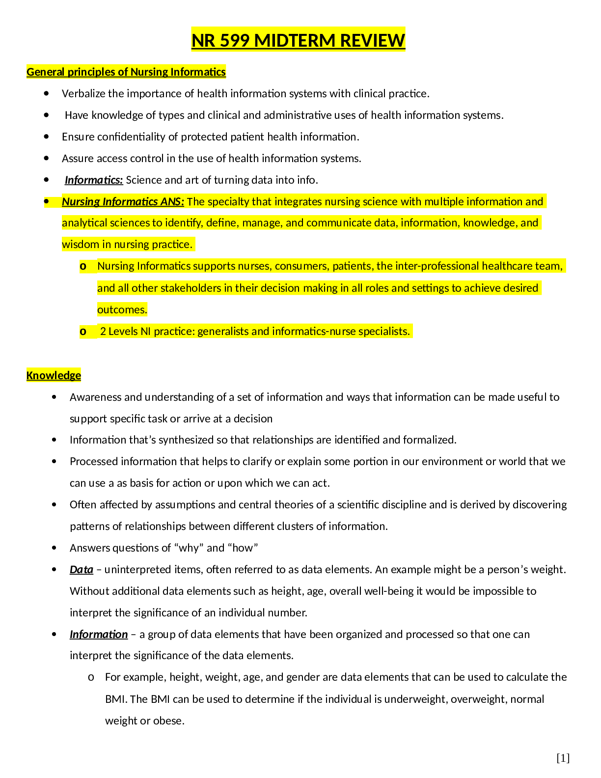
CHAPTER 31- Structure and Function of the Cardiovascular and Lymphatic System A. Circulatory System a. Right Heart Function i. Pumps blood through lungs (pulmonary circulation) ii. Delivers blood ... to the lungs for oxygenation iii. Low pressure system iv. Failures common in IV drug users and back up into lungs b. Left Heart Function i. Pumps oxygenated blood through the systemic circulation ii. Delivers metabolic waste products to the lungs, kidneys and liver iii. High Pressure system iv. Failures back up into the periphery B. The Heart a. Structures that Direct Circulation Through the Heart i. Function 1. Structural Support: includes heart wall and fibrous skeleton which encloses and supports the heart and divides it into four chambers, the valves that direct flow, and the great vessels that conduct blood to and from the heart 2. Maintenance of heart cells: the vessels of coronary circulation- the arteries and veins that serve the metabolic needs of all the heart cells 3. Stimulation and control of heart action: the nerves and specialized muscle cells that direct rhythmic contraction and relaxation, propelling blood throughout circulation ii. Heart wall has 3 layers: 1. Pericardium- outermost, double walled membranous sac that encloses the heart. A surface layer of mesothelium over a thin layer of connective tissue. The visceral layer (epicardium) is the inner layer that folds back on itself and becomes continuous with the parietal pericardium, allowing the large vessels to enter and leave the heart without breaching the pericardial layers. There is a pericardial cavity that separates the layer of the pericardium with 10-30mL of fluid secreted by mesothelium cells to lubricate and minimize friction a. Prevents displacement of the heart during gravitational acceleration or deceleration b. A physical barrier that protects the heart against infection and inflammation from the lungs and pleural space. c. Contains pain receptors and mechanoreceptors that can elicit reflex changes in blood pressure and heart rate 2. Myocardium- the thickest layer of the heart composed of cardiac muscle and is anchored to the fibrous skeleton. Thickness varies from chamber to chamber. Biggest concern for injury to this layer 3. Endocardium- the internal lining of the myocardium composed of connective tissue and a layer of squamous cells. The endocardial lining of the heart is continuous with the endothelium that lines all the arteries, [Show More]
Last updated: 1 year ago
Preview 1 out of 22 pages
.png)
Also available in bundle (1)
Pathophisiology exams with correct answers.
pathophisiology.
By Professor Lynne 2 years ago
$12
5
Reviews( 0 )
Document information
Connected school, study & course
About the document
Uploaded On
May 28, 2021
Number of pages
22
Written in

Additional information
This document has been written for:
Uploaded
May 28, 2021
Downloads
0
Views
61
.png)