Neurology > Lab Report > Chamberlain College of Nursing BIOS 255 Week 4 Lab PART A: Step 1: Analysis of Thymic Tissue an (All)
Chamberlain College of Nursing BIOS 255 Week 4 Lab PART A: Step 1: Analysis of Thymic Tissue and PART B: Step 2: Analysis of Lymphatic Tissue
Document Content and Description Below
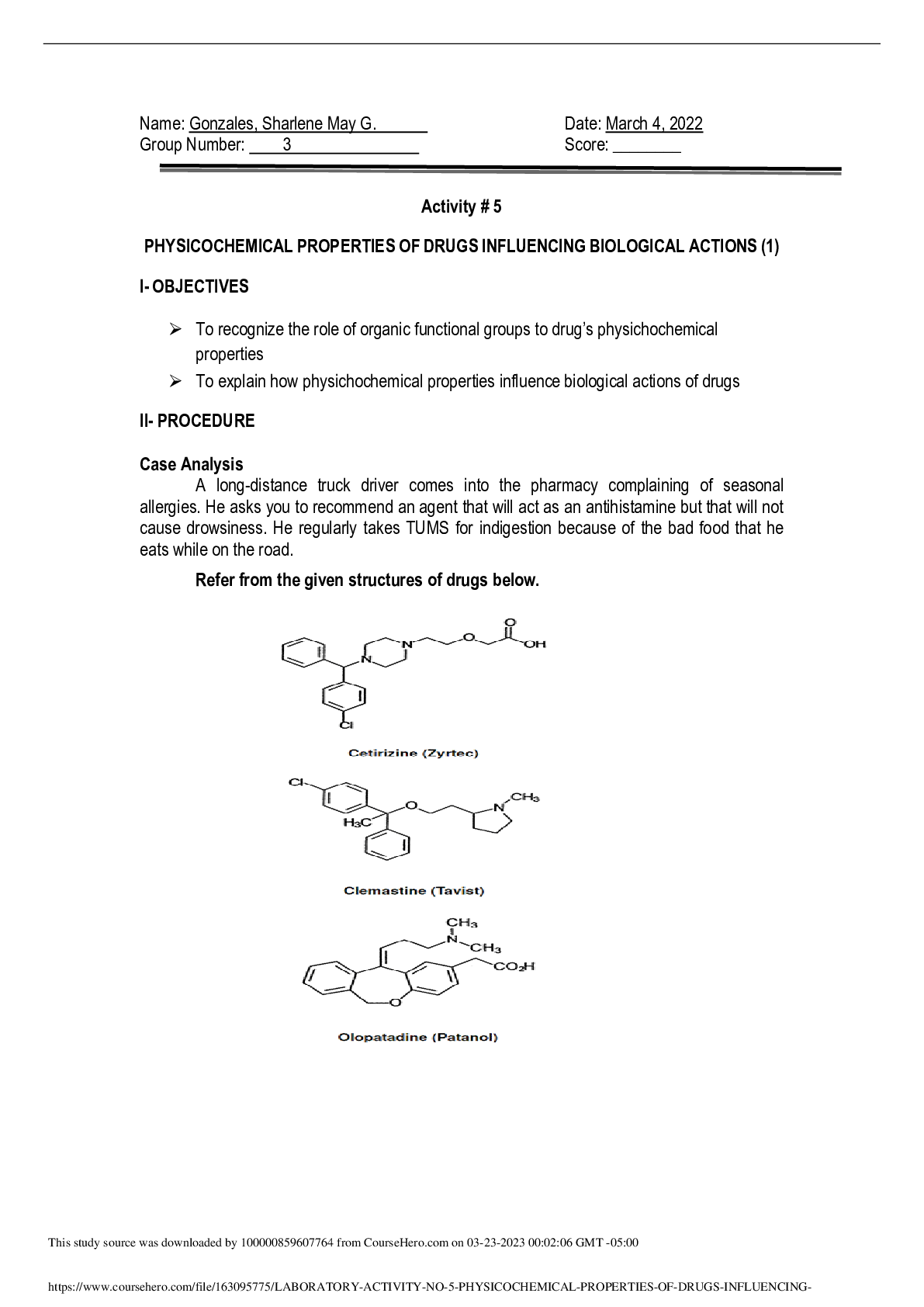
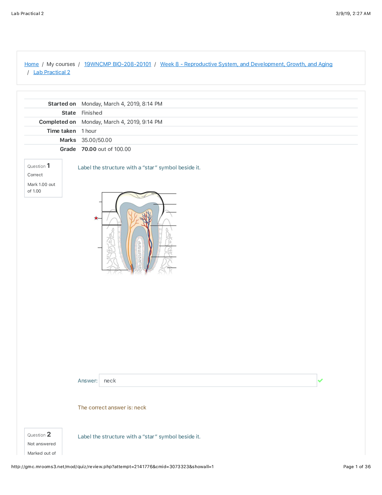
Chamberlain College of Nursing BIOS 255 Week 4 Lab PART A: Step 1: Analysis of Thymic Tissue and PART B: Step 2: Analysis of Lymphatic Tissue Thymus Cortex - The cortex of the thymus... is the outer region of densely packed cells that give rise to T lymphocytes. Thymic corpuscles - are concentrations of flattened epithelial cells in the medulla of the thymus gland. They begin development prior to birth and their numbers increase throughout life. They probably function in removal of dead thymocytes. The medulla of the thymus has few lymphoid cells and contains a variety of non-lymphoid cells that have a neuroendocrine function. Cortex Thymic Corpuscle Medulla T cells T lymphocytes are white blood cells which are produced in the Bone Marrow but matures in the Thymus PART B: Step 2: Analysis of Lymphatic Tissue ~ 1 hour a. Lymphatic cortex (with B cells) The inner cortex of the lymph node consists mainly of T cells and dendritic cells that enter the lymph node from other tissues. The dendritic cells present antigens to T cells, causing their proliferation. The newly formed T cells then migrate from the lymph node to areas of the body where there is antigenic activity. b. Germinal Center This center is a region within the cortex of the lymph node that consists primarily of antigen-stimulated B cells. These are large cells that divide rapidly. c. Lymphatic sinus The medullary sinuses transport the lymph through the medulla of the lymph node. d. Connective tissue capsule 1.2 This is a collagenous connective tissue capsule that surrounds the lymph node and sends connective tissue extensions into the node. PART C: Step 2: Analysis of the Spleenic Tissue ~ ½ hour a. White pulp This is the region of the spleen composed of lymphatic tissue that consists primarily of B-lymphocytes. b. Red pulp This is the portion of the spleen that consists of venous sinuses filled with blood and thin plates of splenic tissue called splenic cords. c. Trabecula This term literally means little beams. The term is used to designate numerous structures in anatomy ranging from ridges of muscle tissue in the heart to extensions of the pia-arachnoid across the subarachnoid space to spicules of bone in spongy bone tissue to fibrous septa of connective tissue within the epithelial masses of certain organs. [Show More]
Last updated: 1 year ago
Preview 1 out of 6 pages
.png)
Reviews( 0 )
Document information
Connected school, study & course
About the document
Uploaded On
Nov 07, 2022
Number of pages
6
Written in

Additional information
This document has been written for:
Uploaded
Nov 07, 2022
Downloads
0
Views
41

.png)

 (1).png)