*NURSING > CASE STUDY > NURS 6501 Knowledge Check Module 7 (All)
NURS 6501 Knowledge Check Module 7
Document Content and Description Below
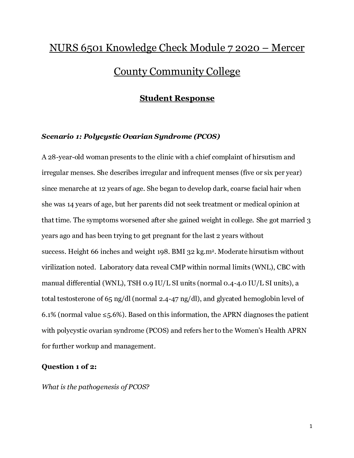
NURS 6501 Knowledge Check Module 7 2022 – Mercer County Community College Student Response Scenario 1: Polycystic Ovarian Syndrome (PCOS) A 28-year-old woman presents to the clinic with a chie... f complaint of hirsutism and irregular menses. She describes irregular and infrequent menses (five or six per year) since menarche at 12 years of age. She began to develop dark, coarse facial hair when she was 14 years of age, but her parents did not seek treatment or medical opinion at that time. The symptoms worsened after she gained weight in college. She got married 3 years ago and has been trying to get pregnant for the last 2 years without success. Height 66 inches and weight 198. BMI 32 kg.m2. Moderate hirsutism without virilization noted. Laboratory data reveal CMP within normal limits (WNL), CBC with manual differential (WNL), TSH 0.9 IU/L SI units (normal 0.4-4.0 IU/L SI units), a total testosterone of 65 ng/dl (normal 2.4-47 ng/dl), and glycated hemoglobin level of 6.1% (normal value ≤5.6%). Based on this information, the APRN diagnoses the patient with polycystic ovarian syndrome (PCOS) and refers her to the Women’s Health APRN for further workup and management. Question 1 of 2: What is the pathogenesis of PCOS? The underlying cause of PCOS is unknown, however a genetic basis is suspected. The pathogenesis of PCOS has been linked to altered luteinizing hormone (LH) action, insulin resistance, and a possible predisposition to hyperandrogenism. One theory maintains that underlying insulin resistance exacerbates hyperandrogenism by suppressing synthesis of sex hormone–binding globulin and increasing adrenal and ovarian synthesis of androgens, thereby increasing androgen levels. These androgens then lead to irregular menses and physical manifestations of hyperandrogenism. The hyperandrogenic state is a cardinal feature of PCOS but glucose intolerance/insulin resistance and hyperinsulinemia often run parallel to and markedly aggravate the hyperandrogenic state, thus contributing to the severity of signs and symptoms of PCOS. Question 2 of 2: How does PCOS affect a woman’s fertility or infertility? Ovulation difficulties are usually the primary cause of infertility in women with PCOS. Ovulation may not occur due to an increase in testosterone production or because follicles on the ovaries do not mature. Due to unbalanced hormones, ovulation and menstruation can be irregular. A hyperandrogenic state is a cardinal feature in the pathogenesis of PCOS. Excessive androgens affect follicular growth, and insulin affects follicular decline by suppressing apoptosis and enabling follicle to persist. There is dysfunction in ovarian follicle development. Inappropriate gonadotropin secretion triggers the beginning of a vicious cycle that perpetuates anovulation. Scenario 2: Pelvic Inflammatory Disease (PID) A 20-year-old female college student presents to the Student Health Clinic with a chief complaint of abdominal pain, foul smelling vaginal discharge, and fever and chills for the past 4 days. She denies nausea, vomiting, or difficulties with defecation. Last bowel movement this morning and was normal for her. Nothing has helped with the pain despite taking ibuprofen 200 mg orally several times a day. She describes the pain as sharp and localizes the pain to her lower abdomen. Past medical history noncontributory. GYN/Social history + for having had unprotected sex while at a fraternity party. Physical exam: thin, Ill appearing anxious looking white female who is moving around on the exam table and unable to find a comfortable position. Temperature 101.6F orally, pulse 120, respirations 22 and regular. Review of systems negative except for chief complaint. Focused assessment of abdomen demonstrated moderate pain to palpation left and right lower quadrants. Upper quadrants soft and non-tender. Bowel sounds diminished in bilateral lower quadrants. Pelvic exam demonstrated + adnexal tenderness, + cervical motion tenderness and copious amounts of greenish thick secretions. The APRN diagnoses the patient as having pelvic inflammatory disease (PID). Question: What is the pathophysiology of PID? Pelvic inflammatory disease (PID) is an infectious and inflammatory disorder of the upper female genital tract, including the uterus, fallopian tubes, or ovaries; in more severe form, the entire peritoneal cavity. The development of upper genital tract infections is mediated by the failure of a number of defense mechanisms that usually are effective in preventing PID. Chlamydia and gonorrhea are the predominant sexually transmitted organism associated with PID. Many anaerobic bacteria have been implicated in increasing the risk of PID because they alter the pH of the vaginal environment and may decrease the integrity of the mucus blocking the cervical canal. Once the infection is established within the uterus and fallopian/uterine tubes, the infection may induce changes in the columnar epithelium lining the upper reproductive tract, causing permanent damage. The resultant inflammatory response causes localized edema and occasionally obstruction or necrosis of the area. Scenario 3: Syphilis A 27-year-old male comes to the clinic with a chief complaint of a “sore on my penis” that has been there for 3 days. He says it burns and leaked a little fluid. He denies any other symptoms. Past medical history noncontributory. Social history: works as a bartender and he states he often “hooks up” with some of the patrons, both male and female after work. He does not always use condoms. Physical exam within normal limits except for a lesion on the lateral side of the penis adjacent to the glans. The area is indurated with a small round raised lesion. The APRN orders laboratory tests, but feels the patient has syphilis. Question: Describe the 4 stages of syphilis Primary: A chancre, or hard lesion develops at the site of the treponemal entry after exposure. In acquired syphilis, T. pallidum rapidly penetrates intact mucous membranes or microscopic dermal abrasions and, within a few hours, enters the lymphatics and blood to produce systemic infection. Incubation time from exposure to development of primary lesions, which occur at the primary site of inoculation, ranges from 12 days to 12 weeks. - - - - - - - - - - - - - - - - - - - - - - - - - - - - - Scenario 14: Thrombotic Thrombocytopenic Purpura (TTP) A 33-year-old female is brought to Urgent Care by her husband who states his wife has gotten suddenly confused and complains of a severe headache. He also noticed large bruises on her legs which were not there yesterday. Only significant past medical history is that the patient developed herpes zoster 2 weeks ago and was given acyclovir for treatment. Physical exam revealed well developed female who is only oriented to person. Large areas of ecchymosis noted on both arms and legs. Stat CBC revealed a platelet count of 18,000/mm3, hemoglobin of 8 g/dl and hematocrit of 24%. The patient was immediately transported to the Emergency Room by Emergency Medical Services (EMS) where further work up demonstrated idiopathic thrombotic thrombocytopenic purpura (TTP). Question: What is the pathophysiology of TTP? TTP characterized by the concomitant occurrence of often severe thrombocytopenia and thrombotic microangiopathy in which platelets aggregate and cause occlusion of arterioles and capillaries within the microcirculation. The underlying mechanism in TTP typically involves antibodies inhibiting the enzyme ADAMTS13. TTP is caused by spontaneous aggregation of platelets and activation of coagulation in the small blood vessels. Platelets are consumed in the aggregation process and bind von Willebrand Factor (vWF). These platelet-vWF complexes form small blood clots which circulate in the blood vessels and cause shearing of red blood cells, resulting in their rupture and formation of schistocytes. These microemboli cause severe organ damage by reducing blood flow to the organ. Scenario 15: Heparin Induced Thrombocytopenia (HIT) A 64-year man is recovering from a transurethral resection of the prostate for treatment of benign prostate hyperplasia. The patient is receiving intravenous antibiotics for the urinary tract infection that was found on the preoperative urine culture and sensitivity (C & S). The post-operative course has been smooth and the APRN is removing the 3-way Foley catheter when there is a sudden release of bright red blood with many blood clots in the Foley bag. The patient becomes hypotensive, tachycardic and the APRN notes new ecchymoses on the patient’s arms and legs. The patient was immediately transferred to the surgical intensive care unit (SICU) and a stat hematology consult was conducted. Stat CBC, d-dimer, peripheral blood smear, partial thromboplastin time, Prothrombin time/international normalization ratio (INR), and fibrinogen labs were drawn. Results were: CBC with markedly decreased platelet count, peripheral blood smear showed decreased number of platelets and presence of large platelets and fragmented red cells (schistocytes), prothrombin time prolonged as was the partial thromboplastin time. The d-dimer was markedly elevated, and fibrinogen level was low. The diagnosis of disseminated intravascular coagulation (DIC) was made based on clinical picture and laboratory data. Question 1 of 2: What is DIC and how does it develop? Disseminated intravascular coagulation (DIC) is characterized by systemic activation of blood coagulation, which results in generation and deposition of fibrin, leading to microvascular thrombi in various organs and contributing to multiple organ dysfunction syndrome (MODS). Consumption of clotting factors and platelets in DIC can result in life-threatening hemorrhage. DIC results from abnormally widespread and ongoing activation of clotting. A variety of conditions are associated with DIC. The common pathway for DIC appears to be excessive and widespread exposure to TF. Wide spread damage to the vascular endothelium results in exposure of subendothelial TF. Question 2 of 2: What factors contribute to the development of DIC? Diagnostic criteria have been established and include a systemic thrombohemorrhagic disorder with laboratory evidence of: clotting activation, fibrinolytic activation, coagulation inhibitor consumption, and biochemical evidence of end-organ damage or failure. DIC is secondary to a wide variety of well-defined clinical conditions, specifically those capable of activating the clotting cascade. These include: arterial hypotension; frequently accompanying shock, hypoxemia, acidemia, and stasis of capillary blood flow. Sepsis is the most common condition associated with DIC. Gram negative/positive microorganisms, fungi, protozoa (malaria), and viruses (influenza, herpes) are capable of precipitating DIC by causing damage to vascular endothelium. [Show More]
Last updated: 1 month ago
Preview 1 out of 22 pages
Reviews( 2 )

by lrussell · 3 years ago
Thanks for the rating by Martin Freeman. 3 years ago

by Martin Freeman · 3 years ago
Document information
Connected school, study & course
About the document
Uploaded On
Nov 12, 2020
Number of pages
22
Written in
Additional information
This document has been written for:
Uploaded
Nov 12, 2020
Downloads
1
Views
70

