*NURSING > STUDY GUIDE > NR 507 Final Study Guide Spring 2020 | NR507 Final Complete Study Guide (All)
NR 507 Final Study Guide Spring 2020 | NR507 Final Complete Study Guide
Document Content and Description Below
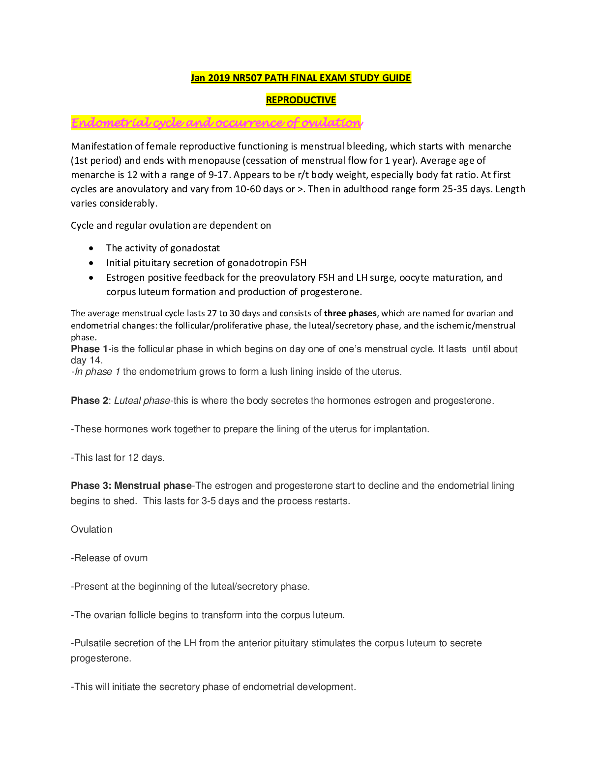
NR 507 Final Study Guide Spring 2020 • Reproductive: o Endometrial cycle (menstrual cycle) and the occurrence of ovulation During menstruation (menses), the functional layer if endometrium ... disintegrates and is discharges through the vagina. Follicular/proliferative phase - GnRH and a balance between activin and inhibin from the granulosa cells contribute to the rise of FSH levels, which stimulates a number of follicles. The pulsatile secretion of FSH from the anterior pituitary gland rescues a dominant ovarian follicle from apoptosis by days 5 to 7 days of the cycle. Together estrogen and FSH increase FSH receptors in the granulosa cells of the primary follicle, making them more sensitive to FSH. FSH and estrogen combine to induce production of LH receptors on the granulosa cells of the primary follicle, thus promoting LH stimulation to combine with FSH stimulation, causing more rapid secretion of follicular estrogen. As estrogen levels increase, FSH levels drop because of an increase in inhibin-B secreted by the granulosa cells in the dominant follicle. This drop in FSH level decreases the growth of the less-developed follicles. Estrogen causes cells of the endometrium to proliferate and stimulates production of LH. Luteal/secretory phase – ovulation marks the beginning of this phase. The ovarian follicle begins its transformation into a corpus luteum. Pulsatile secretion of LH from the anterior pituitary stimulates the corpus luteum to secrete progesterone, which in turn initiates the secretory phase of endometrial development. Glands and blood vessels in the endometrium branch and curl throughout the functional layer, and the glands begin to secrete a thin glycogen- containing fluid, the secretory phase. If conception occurs, the nutrient-laden endometrium is ready for implantation. Human chorionic gonadotropin (HCG) is secreted 3 days after fertilization by blastocytes and maintains the corpus luteum once implantation occurs at about day 6 or 7. HCG can be detected in maternal blood and urine 8 to 10 days after ovulation. Ischemic/menstrual phase • The production of estrogen and progesterone continues until the placenta can adequately maintain hormonal production. If conception and implantation do not occur, the corpus luteum degenerates and ceases production of progesterone and estrogen. Without progesterone or estrogen to maintain it, the endometrium becomes ischemic and disintegrates. Menstruation then occurs marking the beginning of another cycle. Ovulatory cycles appear to have a minimum length of 24 to 26.5 days: the primary ovarian follicle requires 10 to 12.5 days to develop, and the luteal phase appears relatively fixed at 14 days (+/- 3 days). Menstrual blood flow usually lasts 3-7 days, but it may last as long as 8 days or stop after 1 to 2 days and still be considered within normal limits. Ovulation – the release of an ovum from a mature follicle and marks the beginning of the luteal/secretory phase. o Uterine prolapse Descent of the cervix or entire uterus into the vaginal canal. In severe cases, the uterus falls completely through the vagina and protrudes from the introitus. Symptoms of other pelvic floor disorders also may be present. Tx: • Urinary: sensation of incomplete emptying of the bladder, urinary incontinence, urinary frequency/urgency, bladder “splinting” to accomplish voiding • Bowel: constipation or feeling of rectal fullness or blockage, difficult defecation, stool or flatus incontinence • Urgency: manual “splinting” of posterior vaginal wall to accomplish defecation • Pain & Bulging: vaginal, bladder, rectum; pelvic pressure, bulging, pain, lower back pain • Sexual: dyspareunia, decreased sensation, lubrication, arousal • Kegel exercises • Estrogen to improve tone and vascularity of fascial support • Pessary • Weight loss • Avoidance of constipation o Polycystic ovarian syndrome Most common cause of anovulation and ovulatory dysfunction in women. Defined as having at least two of the following three features: irregular ovulation, elevated levels of androgens (testosterone), and the appearance of polycystic ovaries on ultrasound. Polycystic ovaries do not have to be present to diagnose PCOS, and conversely their presence alone does not establish the diagnosis. Initial identification of genes involved in steroid biosynthesis, androgen biosynthesis, and insulin receptors within the ovary indicates genetic involvement. A hyperandrogenic state is a cardinal feature in the pathogenesis of PCOS. However, glucose intolerance/insulin resistance and hyperinsulinemia often run parallel to and markedly aggravate the hyperandrogenic state, thus contributing to the severity of signs and symptoms of PCOS. Excessive androgens affect follicular growth, and insulin affects follicular decline by suppressing apoptosis and enabling follicles to persist. Weight gain tends to aggravate symptoms, whereas weight loss may ameliorate some of the endocrine and metabolic events and thus decrease symptoms. Women with PCOS tend to have increased leptin levels. Leptin influences the hypothalamic pulsatility of GnRH and consequent interaction along the entire HPO axis. In PCOS there is dysfunction in ovarian follicle development. Inappropriate gonadotropin secretion triggers the beginning of a vicious cycle that perpetuates anovulation. Typically, levels of FSH are low or below normal and LH levels and LH bioactivity are elevated. An increased frequency of GnRH pulses appears to cause increased frequency of LH pulses. Persistent LH elevation causes a increase in the levels of androgens. Androgens are converted to estrogen in peripheral tissues, and increased testosterone levels cause a significant reduction in SHBG, which in turn causes increased levels of free estradiol. Elevated estrogen levels trigger a positive-feedback response in LH and a negative-feedback response in FSH. The accumulation of follicular tissue is various stages of development allows an increased and relatively constant production of steroids in response to gonadotropin stimulation. Thus PCOS is characterized by excessive production of both androgen and estrogen. In turn, persistent anovulation causes enlarged polycystic ovaries characterized by a smooth, pearly white capsule. This characteristic appearance is caused by an increase of surface area and increased volume of up to 2.8 times, doubling of growing and atretic follicles, thickening of the tunica by 50%, increasing cortical stromal thickening by one-third and a fivefold increase in subcortical stroma, and escalating hyperplasia. Manifestations: • Usually appear within 2 years of puberty but may present after a variable period of normal menstrual function and possibly pregnancy. • Symptoms are related to anovulation, hyperandrogenism, and insulin resistance and include dysfunctional bleeding or amenorrhea, hirsutism, acne, acanthosis nigricans, and infertility. Eval & Treatment: • Diagnosis is based on evidence of androgen excess, chronic anovulation, and sonographic evidence of polycystic ovaries with at least 2 of the 3 criteria present. • Tests for impaired glucose tolerance are recommended. • Evidence of hyperandrogenism must be present before PCOS is diagnosed in an adolescent female. • Goals of tx: reversing signs and symptoms of androgen excess, instituting cyclin menstruation, restoring fertility, and ameliorating any associated metabolic or endocrine, or both, disturbances. • First line: combined oral contraceptives for management of symptoms and to establish regular menses. • For those women with PCOS who are overweight or obese, lifestyle modifications including regular exercise and weight loss, also are considered first-line treatment. o Testicular cancer and conditions that increase risk Highly treatable, usually curable cancer that most often develops in young and middle-aged men 90% of testicular cancers are germ cell tumors arising from the male gametes. In addition, testicular tumors can arise from specialized calls of the gonadal stroma. These tumors, which are named for their cellular origins are Leydig cell, Sertoli cell, granulosa cell, and theca cell tumors and constitute less than 10% of all testicular cancer. Risk factors: history of cryptorchidism, abnormal testicular development, human immunodeficiency virus (HIV) and AIDS, Klinefelter syndrome, and history of testicular cancer. Manifestations: • Painless testicular enlargement is usually the first sign. • Enlargement is usually gradual and may be accompanied by a sensation of testicular heaviness or dull ache in the lower abdomen. • Lumbar pain may be present and usually is caused by retroperitoneal node metastasis. • Signs of metastasis to the lungs: cough, dyspnea, hemoptysis • Supraclavicular node involvement: dysphagia, neck swelling • Metastasis to CNS: alterations in vision, mental status, papilledema, and seizures o Symptoms that require evaluation for breast cancer The first sign of breast cancer is usually a painless lump. Lumps caused by breast tumors do not have any classic characteristics. Chest pain (lung metastasis) Dilated blood vessels Dimpling of the skin Edema Edema of the arm Hemorrhage Local pain Nipple/areolar eczema Nipple discharge in nonlactating woman Nipple retraction Pitting of the skin (peaud’orange) Reddened skin, local tenderness, and warmth Skin retraction Ulceration o Signs of premenstrual dysphoric disorder >/= 5 symptoms below: occur in most cycles during the week before menses onset, improve within a few days after menses onset, and diminish in the week postmenses • Marked affective lability • Marked irritability or anger or increased interpersonal conflicts • Marked anxiety, tension • Decreased interest • Difficulty concentrating • Easy fatigability, low energy • Increase or decrease in sleep • Feelings of being overwhelmed • Physical symptoms: breast tenderness, muscle or joint aches, “bloating” or weight gain o Dysfunctional uterine bleeding (Abnormal uterine bleeding) Bleeding that is abnormal in duration, volume, frequency, or regularity and has been present for the majority of the previous 6 months. May be acute or chronic and is classified by PALM-COEIN system: • Polp • Adenomyosis • Leiomyoma • Malignancy • Hyperplasia • Coagulopathy • Ovulatory dysfunction • Endometrial • Iatrogenic • Not-yet classified In premenstrual or menopausal women, any bleeding is considered abnormal. Therefore bleeding more frequently than every 21 days or less frequently than every 35 days, is considered to be abnormal. Menstrual bleeding for longer than 7 days also is considered abnormal. AUB is the leading reason for hysterectomy. Perimenopausal women are most commonly affected. The majority of AUB is due to lack of ovulation. Normal, regular periods are the result of complex interplay between the hypothalamus, pituitary, ovary, and the uterine endometrium. Disruptions in this system can affect the amount and structure of the uterine endometrium, causing it to shed irregularly or heavily. If a follicle forms but never releases the ovum, the follicle may continue to produce estrogen, encouraging endometrial proliferation beyond the normal 14-day time window. In addition, the lack of progesterone causes the thickened endometrium to be unable to shed in a predictable fashion without excessive blood loss. Women who fail to ovulate experience irregularities in their menstrual bleeding related to the lack of progesterone and, in some cases, an excess of estrogen. Without ovulation, menstrual flow may become irregular, excessive, or both, resulting from the large quantity of tissue available for bleeding and the random breakdown of tissue that results in exposure of vascular channels. In the absence of adequate progesterone levels, usual endometrial control mechanisms are missing, such as vasoconstrictive rhythmicity, tight coiling of spiral vessels, and orderly collapse, and stasis does not occur. AUB also can result from defects of the corpus luteum, resulting in progesterone deficiencies, or from abnormalities of the uterus or cervix, such as endometrial polyps, uterine fibroids, or even uterine or cervical cancers. Coagulation defects also can cause heavy and abnormal uterine bleeding and should be suspected in younger women with a history of extensive bruising or bleeding during dental procedures. Iatrogenic AUB can be caused by intrauterine devices or long-acting contraceptive implants or medications, such as anticoagulants, steroids, digitalis, phenytoin, or hypothalamic depressants. Manifestations: [Show More]
Last updated: 1 year ago
Preview 1 out of 45 pages
Instant download

Buy this document to get the full access instantly
Instant Download Access after purchase
Add to cartInstant download
Reviews( 0 )
Document information
Connected school, study & course
About the document
Uploaded On
Apr 17, 2021
Number of pages
45
Written in

Additional information
This document has been written for:
Uploaded
Apr 17, 2021
Downloads
0
Views
25