Chemistry > Study Notes > CHEM120 Week 7 Concepts: Biochemistry – Download For Revision And Improve Your Grades (All)
CHEM120 Week 7 Concepts: Biochemistry – Download For Revision And Improve Your Grades
Document Content and Description Below
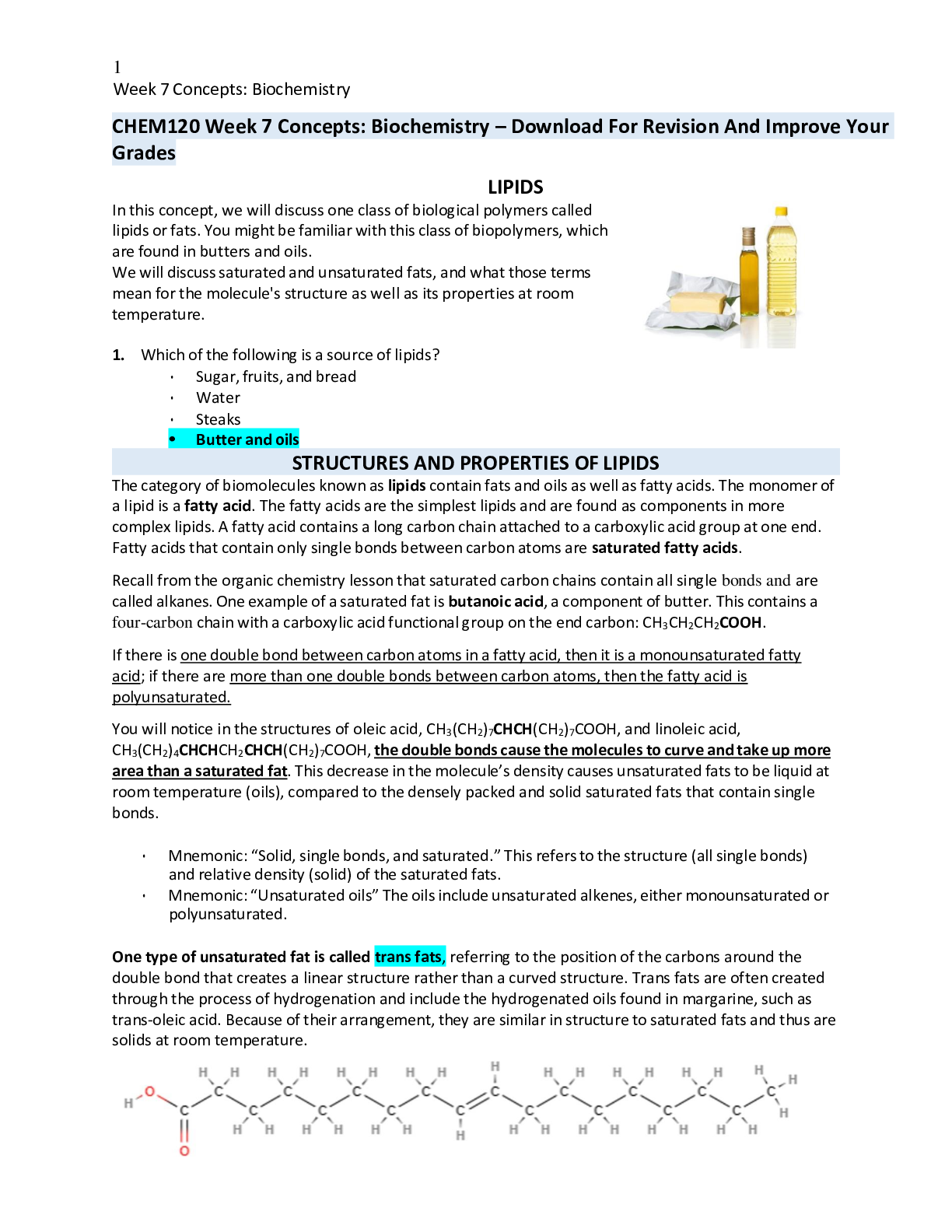
CHEM120 Week 7 Concepts: Biochemistry – Download For Revision And Improve Your Grades LIPIDS In this concept, we will discuss one class of biological polymers called lipids or fats. You might be f... amiliar with this class of biopolymers, which are found in butters and oils. We will discuss saturated and unsaturated fats, and what those terms mean for the molecule's structure as well as its properties at room temperature. 1. Which of the following is a source of lipids? • Sugar, fruits, and bread • Water • Steaks • Butter and oils The category of biomolecules known as lipids contain fats and oils as well as fatty acids. The monomer of a lipid is a fatty acid. The fatty acids are the simplest lipids and are found as components in more complex lipids. A fatty acid contains a long carbon chain attached to a carboxylic acid group at one end. Fatty acids that contain only single bonds between carbon atoms are saturated fatty acids. Recall from the organic chemistry lesson that saturated carbon chains contain all single bonds and are called alkanes. One example of a saturated fat is butanoic acid, a component of butter. This contains a four-carbon chain with a carboxylic acid functional group on the end carbon: CH3CH2CH2COOH. If there is one double bond between carbon atoms in a fatty acid, then it is a monounsaturated fatty acid; if there are more than one double bonds between carbon atoms, then the fatty acid is polyunsaturated. You will notice in the structures of oleic acid, CH3(CH2)7CHCH(CH2)7COOH, and linoleic acid, CH3(CH2)4CHCHCH2CHCH(CH2)7COOH, the double bonds cause the molecules to curve and take up more area than a saturated fat. This decrease in the molecule’s density causes unsaturated fats to be liquid at room temperature (oils), compared to the densely packed and solid saturated fats that contain single bonds. • Mnemonic: “Solid, single bonds, and saturated.” This refers to the structure (all single bonds) and relative density (solid) of the saturated fats. • Mnemonic: “Unsaturated oils” The oils include unsaturated alkenes, either monounsaturated or polyunsaturated. One type of unsaturated fat is called trans fats, referring to the position of the carbons around the double bond that creates a linear structure rather than a curved structure. Trans fats are often created through the process of hydrogenation and include the hydrogenated oils found in margarine, such as trans-oleic acid. Because of their arrangement, they are similar in structure to saturated fats and thus are solids at room temperature. SATURATED OR UNSATURATED Lipids UNSATURATED SATURATED CH3CHCHCH2CHCHCH2COOH CH3CH2CH2CH2CH2COOH CH2CH2CH2COOH CH3CH2CH2CHCHCHCHCH2CH2COOH Answer the following questions about lipids 2. Triglycerides are: • Always unsaturated • Made of fatty acid monomers • Used for catalysis in the body • Soluble in water Select all that apply. 3. Saturated fatty acids contain functional groups • Alkanes • Amine • Carboxylic acid • Halogen • Alkenes From the condensed structural formula, determine if the structure represents a saturated fatty acid, monounsaturated fatty acid, or a polyunsaturated fatty acid. SATURATED FATTY ACID MONOUNSATURATED FATTY ACID POLYUNSATURATED FATTY ACID CH3CH2CHCHCH2COOH X CH3CHCHCH2CH2COOH X CH3CHCHCH2CHCHCH2COOH CH3CH2CH2CH2CH2COOH X 4. Contains carboxylic acid = Both Saturated and Unsaturated Contains a single double carbon-to-carbon bond = Unsaturated Contains multiple double carbon-to-carbon bonds = Unsaturated Solid at room temperature = Saturated Contains no carbon-to-carbon double bonds = Saturated Liquid at room temp = Unsaturated Does not mix with water = Both Saturated and Unsaturated Both saturated and unsaturated do not mix with water. Saturated contains no double carbon-to-carbon bonds and is solid at room temperature. Unsaturated contains a single double carbon-to-carbon bond (monounsaturated) or multiple double carbon-to-carbon bonds (polyunsaturated) and are mostly liquid at room temperature. TRIGLYCERIDES, GLYCEROPHOSPHOLIPIDS, AND STEROID Fats and oils consist of fatty acids that have been bound to glycerol through an ester bond. Lipids, or fats and oils, are also found in abundance in our kitchens. While the carboxylic acid group is soluble in water, the long carbon chain makes the fatty acid insoluble in water. You can observe this by pouring equal amounts of water and vegetable or olive oil into a small, clear container. Two layers will appear: one is the polar (hydrophilic) water, one is the nonpolar (hydrophobic) oil. Because of the differences in hydrophobicity, polar and nonpolar molecules will not mix. This idea is commonly referred to as “Like Dissolves Like,” meaning polar (hydrophilic) molecules will only mix with other polar (hydrophilic) molecules; nonpolar (hydrophobic) molecules will only mix with other nonpolar molecules. Triacylglycerols, or triglycerides, are triesters of glycerol (a trihydroxy alcohol) and fatty acids. Basically, the glycerol molecule has three tails of fatty acids. These molecules are formed by esterification, the condensation of the carboxylic acid on the fatty acid and the alcohol groups on the glycerol. Triacylglycerols also undergo hydrogenation, hydrolysis, and saponification (soap-making) reactions. Glycerophospholipids are similar to triacylglycerols, except that one hydroxyl group of glycerol has been replaced by the ester of phosphoric acid and an amino alcohol and bonded through a phosphodiester bond. One consequence of their structure is that glycerophospholipids have both polar and nonpolar regions. These macromolecules are the most abundant lipid in cell membranes. Cell Membrane lipid bilayer with receptors Steroids are compounds that consist of three cyclohexane rings and one cyclopentane ring that are fused together. This structure is referred to as the steroid nucleus. A very common steroid is cholesterol, which is a component of cellular membranes, myelin sheath tissue, and brain and nerve tissue. Cholesterol is also used to synthesize Vitamin D and steroid hormones. FATTY ACIDS ATTACHING TO GLYCEROL Using the molecule below, answer the following questions 5. This molecule is best described as • A polyunsaturated fatty acid • An unsaturated fatty acid • A triglyceride • A fatty acid 6. How many fatty acids were used to create this triacylglycerol molecule? • One • Two • Three • Four 7. The fatty acids used to create this triacylglycerol are • All unsaturated • All saturated • All connected through an ester linkage • All identical 8. Match the lipid to the function • Glycerophospholipids compromises the majority of cell membranes because it contains a polar head and a nonpolar tail • Triacylglycerol used in soap making • Cholesterol structure included three six-member rings fused with a five-member rings, and in cell membranes provides stability and is a precursor for vitamin D 9. Select all that apply. Which of the following are typically solids at room temperature? • Trans lipids • Saturated lipids • Unsaturated lipids • Monosaturated lipids • Polysaturated lipids 10. Which class of lipids is found in cell membranes? • Sterols • Triacylglycerol • Glycerophospholipid • Steroids and triacylglycerol • Steroids and glycerophospholipids 11. What type of linkage is found in triacylglycerol’s? • Amide • Ether • Peptide • Ester • Glycoside 12. Triacylglycerols contain . • 3 molecules of glycerol and 1 steroid • 1 molecule of glycerol and 3 molecules of fatty acid • 1 molecule of glycerol and 3 steroids • 3 molecules of glycerol and 1 molecule of fatty acid 13. Which of the following is true for both saturated and unsaturated fatty acids? • Both are considered alkenes • Both are solid at room temperature • Both contain ester linkages • Both are soluble in water • Both contain carboxylic acid groups PROTEINS In this concept we discuss the third class of macromolecules: the proteins. Proteins are essential for multiple functions within cells: catalysis, structure, transport, energy, and many others. We will discuss the role of the amide (peptide) linkage, four levels of protein structures, and properties of the proteins. 14. Rank the levels of protein structure from top (least complex/ structured) to bottom (most complex/ structured) • Primary – sequence of amino acids • Secondary – alpha helices, beta strands, and loops • Tertiary – the three-dimensional structure • Quaternary – interaction of multiple tertiary structures 15. Examples of proteins include • Meat and muscle • Sugars and candy • Butter and oil • All of these STRUCTURE OF AMINO ACIDS As with our other biomolecules, to understand proteins, we first need to understand their parts. Proteins are polymers of amino acids. Amino acids have two functional groups: an amino group (-NH2) and a carboxylic acid group (-COOH). This is where their name comes from: amino acid. For all amino acids found in proteins, the amino group, the carboxylic acid group, and a hydrogen atom are bonded to the central carbon atom (called the alpha carbon). The R is the placeholder for the sidechain. Different amino acids have different side chains, but they all have the amine group, alpha carbon, and carboxylic acid groups. While there are many amino acids, only 20 different amino acids are found in humans. On the general structure of an amino acid below, click on the following parts: amino group, carboxylic acid group, and alpha hydrogen. General structure of an amino acid can be written as NH2CHRCOOH. The backbone is the portion that repeats in every amino acid: the amine, the alpha carbon, and the carboxylic acid. To show a specific amino acid, the R is replaced by the sidechain. One of the twenty amino acids is shown below. As we can see, we have replaced "R" with a -CH3. This -CH3 is one of many possible side chains. The name of this particular amino acid is Alanine and it is one of the 20 amino acids found in our bodies. Two amino acids will join when the –OH from the carboxylic acid of the first molecule condenses with the amine group of the next amino acid, releasing a molecule of water as a by-product. AMINO ACID SIDE CHAINS 16. The backbone of an amino acid refers to all of the following except • The alpha carbon • The amine • The carboxylic acid • The sidechain 17. Select all that apply. What functional groups are found in amino acids? • Carboxylic acid • Ester • Amine • Alkene • Hydroxyl 18. Alpha carbons are always bound to • the amine. • a hydrogen. • the carboxylic acid. • the sidechain. • All of these. Two amino acids will join when the –OH from the carboxylic acid of the first molecule condenses with the amine group of the next amino acid, releasing a molecule of water as a by-product. A chain of two amino acids (like the one we formed above) is a dipeptide. A chain of many amino acids is a polypeptide. When there are more than 50 amino acid residues in a polypeptide, it is usually called a protein. A peptide is drawn from the amino terminus (NH2-) to the carboxyl terminus (-COOH), or from the N-terminus to the C-terminus. This is the order of the protein sequence. You can remember by remembering it is the same order as the name of the monomer: amino acid. The sequence starts on the amine (N) and ends on the acid (COOH). On the image below of the dipeptide we just formed, click on the N-terminus and the C-terminus. DIPEPTIDE Locate these parts of the dipeptide: Peptide Bond, N-Terminus and C-Terminus 19. Protein sequences go from the N- terminus to the C- terminus. The N terminus represents the first amino group of the protein backbone, and the C terminus represents the last carboxylic acid group of the protein backbone. LEVELS OF PROTEIN STRUCTURE The structure of a polypeptide or protein is essential for its function. Below, we explore the structural levels of protein structure. When referring to peptides, the polypeptide is depicted as a chain of amino acids, each represented by their three-letter abbreviation. This is sometimes called the protein sequence. For example, the polypeptide that consists of glycine, alanine, and serine is shown as Gly-Ala-Ser. This order of amino acids is called the primary structure and is the code of the polypeptide or protein. • Primary structure – sequence of amino acids held together by peptide bonds. The order of amino acids will determine all following levels of structure. • Secondary structure – the way that the local amino and carboxylic acid groups interact with each other in space, mostly through hydrogen bonding interactions that are nearby interactions (alpha helices), distant interactions (beta strands) or no interactions (loops). This is based on the sequence of amino acids in the primary structure. • Tertiary structure – the overall, three-dimensional and completely folded structure. This is largely due to hydrophobic sidechains burying themselves away from surrounding water, though other interactions also play a part. This is an overall bending of the secondary sequence at the loops, pushing the secondary structures into a 3D structure. • Quaternary structure – the combination of two or more tertiary protein structures to form more complex and functional proteins. Not all proteins require this level to be functional. Covalent bonds, specifically amide (peptide) bonds, are the driving force of primary structure. The covalent bonds are made through polymerization (condensation) and can only be broken in a chemical reaction such as hydrolysis. All other levels of structure are primarily driven by intermolecular forces that form spontaneously due to their molecular properties, particularly polarity. See the table below for more information about protein structure. LEVEL OF STRUCTURE DEFINITION PRIMARY INTERACTIONS EXAMPLES Primary Order of amino acids in the peptide Amide (peptide) bonds joining the amino acids Gly-Ala-Ser Secondary The way amino acids lie next to each other in space Hydrogen bonding between the acid -CO- and the amine -NH- in the primary sequence backbone • Alpha helix (close hydrogen bonds) • Beta strand (distant hydrogen bonds) • Loop (flexible regions with no hydrogen bonds) Tertiary Overall 3D shape of a protein chain • Hydrophobic (most important) • Hydrophilic • Hydrogen bonds • Disulfide bonds • Salts bridges • Dispersion forces Globular structure of my myoglobin Quaternary Interaction of multiple tertiary protein chains • Hydrophobic • Hydrophilic • Dispersion forces • Salt bridges There are four protein chains that interact to form the active, quaternary structure of hemoglobin LEVELS OF PROTEIN STRUCTURE: AN ANALOGY The structure levels of a polypeptide or protein can be likened to written communication. The primary structure is like the order of letters: thecatisneverfarfromfood The secondary structure is analogous to how we could sort the nearby letters to form words: the cat is never far from food The tertiary structure is how these words interact to form a sentence, giving us a thought when we see how each of the words interacts: The cat is never far from food. Finally, like quaternary structure, sometimes we need more than one sentence to explain a situation. A quaternary structure is like how we can use multiple sentences to form a paragraph from several complete sentences, each with a beginning and an end: The cat is never far from food. She jumps on the table whenever I eat. How can she be so hungry all the time if she spends all day sleeping? 20. Click and drag to match the structure to the description LEVEL OF STRUCTURE DESCRIPTION PRIMARY sequence of amino acids held together by peptide bonds SECONDARY the way that the local amino and carboxylic acid groups interact with each other in space TERTIARY the overall, three-dimensional and completely folded structure QUATERNARY the combination of two or more tertiary protein structures 21. What is the driving force of each level of structure? Peptide bonds H-bonding of the backbone Hydrophobic sidechain interactions Primary X Secondar y X Tertiary X If a protein’s structure is disrupted, it will no longer be able to function. Protein denaturation occurs when the environment changes and the protein’s secondary, tertiary, and quaternary structures are disrupted. High pH (basic), low pH (acidic), high salt, or high temperature can disturb the intermolecular forces that drive those levels of structure. The environment typically does not affect the peptide bonds involved in the primary structure, and occasionally the protein can re-fold but it will never be functional again. You have likely seen this if you have ever observed a cooked egg: the proteins in the egg whites start as clear and gooey, but become white and solid when high temperature is added. Once cooked, it cannot be re-formed into an uncooked egg. When lemon juice, an acid, is added to milk the proteins in milk will become solid curds at this change in pH. Because proteins play many essential roles in cells the pH, temperature, and salt concentration are carefully maintained in human bodies to ensure that the cells can function. Without close regulation, the proteins in the cell would denature and the cell would perish. Complete the following activity and then answer the questions below about protein structure. 1. Locate two long pieces of thin, bendable wire in your home (such as pipe cleaner, garden wire, electrical wire, or similar), preferably 5-10 inches in length as well as a pencil. 2. Straighten the wire and while grabbing each end, give it a good tug. This represents the primary structure, held together with covalent bonds. 3. Next, twirl 2 inches of the wire around a pencil to make a spiral, leave a small gap of 1-2 inches of straight wire, then crinkle the next part two inches; the model should be in a relatively straight line as you complete this process. Repeat this pattern, or any pattern, for the remaining length of the wire. This represents the secondary structure elements: alpha helix is the spiral, the loop is unstructured, and the crinkles are the beta strands. This structure is held in place by hydrogen bonds, intermolecular forces that are not as strong as covalent bonds. 4. Next, bend the structure at the loops to bury the hydrophobic sidechains and make a 3D structure. This represents the tertiary structure, in which hydrophobic sidechains are buried, hydrophilic sidechains are left on the surface, and various other intermolecular interactions occur to lock the 3D structure in place: disulfide bridges form between two sulfur-containing sidechains; salt bridges form between cationic and anionic sidechains, and hydrogen bonds between polar Hydrogen and nitrogen or oxygen form. Some proteins will be fully functional at the tertiary structure, but we will model the quaternary structure. 5. Finally, take the second wire and repeat the process. When you have your two complete tertiary structures, place them near to each other to represent the quaternary structure, which is made from two or more tertiary sequences. Next, we will model denaturation. In this process, a protein will lose most of its structure due to high salt, high temperature, a low pH, or a high pH. When a protein loses its structure, it loses the ability to function. Salt, temperature, and pH will cause the intermolecular forces to become interrupted. Move the two protein chains apart to denature the quaternary structure. Pick up one chain and holding both ends as you did when starting the demo, pull the ends to straighten the wire. 22. What level of structure did the straightened wire represent throughout the demonstration? • primary • secondary • tertiary • quaternary 23. How did secondary structure differ from tertiary structure in this demo? • secondary structure was the straight wire, tertiary structure was the twists and crinkles • secondary structure was when the straightened wire was twisted and crinkled, tertiary was when the twists and crinkles were bent further to make a 3D shape • secondary structure and tertiary structure were indistinguishable in this demo • secondary structure is the completed 3D protein chain, tertiary structure was when the second chain was placed next to the first chain. 24. After denaturation, what level of protein structure remained? • primary • secondary • tertiary • quaternary 25. Select all that apply. What levels or protein structure were affected by denaturation? • primary • secondary • tertiary • quaternary 26. What causes denaturation? Select all that apply. • high pH • low pH • high salt • high temperature 27. The primary structure of a protein is held together by • peptide bonds. • glycosidic bonds. • ester bonds. • All of these 28. Which level of protein structure is the three-dimensional shape? • Primary • Secondary • Tertiary • Quaternary 29. Which of the following is unique to each amino acid? • The amine • The alpha carbon • The carboxylic acid • The sidechain 30. Select all that apply. What is the result when a protein is denatured? • the primary structure is altered • the secondary structure is altered • the tertiary structure is altered • the quaternary structure is altered • all four levels of protein structure are altered • it can no longer function 31. identify the sidechain in the dipeptide • A • B C • D 32. What causes denaturation? • high pH • low pH • high salt • high temperature • all of the above • none of the above 33. Which level of protein structure is the interaction of multiple protein chains? • primary • secondary • tertiary • quaternary ENZYMES In this concept we discuss a special class of proteins called enzymes, which perform chemical reactions in the cell. These proteins help us get energy from food through catabolism, as well as build new molecules through anabolism. We will discuss the specific terminology related to enzymes as well as their roles. 34. When an enzyme is denatured, this will affect both its structure and its function. • True • False, this will not affect its structure. • False, this will not affect its function. • False, this will not affect either structure or function. 35. Which of the following macromolecules best describes enzymes? • carbohydrates • proteins • lipids • nucleic acids Enzymes are a type of protein that performs chemical reactions. The enzymatic action of proteins is very interesting because enzymes serve as a catalyst in a reaction to lower the activation energy for the reaction. Catalysts increase the speed of the reaction without being consumed in the reaction. This lowered activation energy allows the chemical reactions to happen faster – on the same timescale organisms need to live and grow. Without enzymes, these chemical reactions would take years. The reactant molecule an enzyme acts on is known as the substrate. The substrate fits into the active site of the enzyme where the reaction occurs. Finally, the product(s) are released by the enzyme. As an important note, each enzyme only acts on a single substrate or a closely related group of substrates. This is due to the size and polarity of the active site: only certain molecules will fit into the active site. One example of an enzyme is alcohol dehydrogenase. This enzyme uses ethanol as its substrate. The substrate ethanol binds the active site in the enzyme, shown by an arrow in the image. The flexible enzyme closes over the substrate to make the enzyme-substrate complex that specifically positions the substrate for a quick reaction. Once in position, the substrate reacts to become the products, Ethan aldehyde and H+. When the chemical reaction is over, the products, ethanal and H+, are released from the active site. The empty enzyme is then able to bind another molecule of ethanol, allowing the process to continue as long as the enzyme’s structure is maintained. In the activity below, click to identify the active site, substrate, and product in the image of alcohol dehydrogenase. Answer the questions below about the structure and role of enzymes. 36. An ENZYME is a type of protein that performs chemical reactions quickly without being consumed, also called a CATALYST. To start the reaction, the enzyme must bind the SUBSTRATE molecule in its ACTIVE SITE, a specific location in the enzyme where the chemical reaction will occur. When the chemical reaction is complete, the PRODUCT is released, and the enzyme can start the process again. 37. True or false. Enzymes bind a single substrate or class of substrates. • True • False 38. Which part of the enzyme allows substrate specificity? • reaction site • protein • active site • allosteric site FACTORS AFFECTING ENZYME ACTIVITY Enzymes play key roles in metabolism: the building (catabolic) and breaking (anabolic) reactions done daily in cells. Enzymes are responsible for catabolizing nutrients into building blocks, and then using these building blocks to anabolize the polymers in our bodies, including glycogen for energy storage, proteins for structure, and even DNA for information. There are numerous examples of both catabolism and anabolism. Any time a protein’s structure is denatured it can no longer function. Enzymes are one type of protein, so they, too, can be denatured in high temperature, high salt, high pH, or low pH. The body carefully maintains a regular temperature, pH, and salt concentration in order to optimize enzyme function throughout the body. Some enzymes even function in the low pH of your stomach and will become disabled in the high pH of the intestines. The structures are adapted to the environment in which they are active. Some enzymes require cofactors or coenzymes to complete their tertiary or quaternary structure. Cofactors are inorganic ions, such as iron in hemoglobin. Coenzymes are organic vitamins. When a cofactor or coenzyme is present in the structure, the complete structure is called the holoenzyme. When missing, the protein-only structure is called the apoenzyme (“apo” meaning “without”) and is not functional. Inhibitors stop an enzyme from functioning by either altering its tertiary structure or blocking the active site. Both of these prevent the substrate from binding to the active site. Competitive inhibition, a molecule that greatly resembles the substrate will bind the active site. Competitive inhibition will not affect protein structure, and competitive inhibitors can be removed easily to start catalysis again. In noncompetitive inhibition, a molecule will bind a different site on the enzyme away from the active site, called the allosteric site; this will change the tertiary structure to alter the shape or even fully close the active site. When the active site is altered in this way, the substrate cannot bind, and the reaction cannot proceed. FACTOR THAT AFFECTS ENZYMES WHY EXAMPLES Denaturing This disrupts protein shape, thus disrupting its function • High pH • Low pH • High temp • High Salt Cofactors and coenzymes In some proteins, these molecules are required to maintain the structure of the enzyme • Cofactors are inorganic metals (Fe2+, Ca2+, Mg2+, etc) • Coenzymes are organic vitamins (niacin, folic acid, etc) Inhibitors Inhibitors will stop the substrate from binding, thus stopping the reaction • Competitive inhibition: a molecule similar to the substrate competes for the active site and binds instead • Noncompetitive inhibition: a molecule binds to an allosteric site, which causes the enzyme to change shape and close the active site Answer the following questions below about factors that affect enzyme activity 39. Which of the following affects an enzyme’s ability to function? • High pH • Low pH • Cofactors • Inhibitors • All of these above affect an enzyme’s ability to function 40. A molecule that mimics the substrate in size and polarity binds the active site, preventing the true substrate from being catalyzed. This is an example of • noncompetitive inhibition. • a cofactor. • competitive inhibition. • a coenzyme. • denaturing. 41. A molecule binds an allosteric site on the enzyme away from the active site, which causes the enzyme’s active site to change shape. This is an example of • a coenzyme. • noncompetitive inhibition. • competitive inhibition. • a cofactor. • denaturing. There are several ways that enzyme structure and function are affected. Mark whether the example will affect the protein’s tertiary structure and overall folding, or if it will not affect the protein’s structure. Affects enzyme’s tertiary structure Does not affect enzyme’s tertiary structure Coenzymes X Competitive inhibition X Noncompetitive inhibition X Denaturing X 42. When , inorganic ions required for the complete structure of the enzyme, are not incorporated into the enzyme structure, the remaining protein-only portion is called . • cofactor / holoenzyme • coenzyme / apoenzyme • cofactor / apoenzyme • coenzyme / holoenzyme 43. Determine if the example would act as a coenzyme or a cofactor in a holoenzyme. Coenzyme Cofactor Vitamin B12 X Iron X Niacin X Magnesium X Calcium X 44. Which of the following is an example of a typical enzyme function? • Catabolizing nutrients into monomer building blocks • Anabolizing glycogen • Catalyzing the construction of DNA • All of these 45. Enzymes decrease • the speed of a reaction. • the number of products in a reaction. • the total number of reactions. • the activation energy of a reaction. 46. How many substrates does an enzyme typically have? • One, or one group of similar substrates • Two, or two groups of similar substrates • It varies, but typically one for each quaternary structure • Many: there is no limit 47. Which of the following is incorrectly matched? • cofactor – inorganic ion required for holoenzyme function • activation energy – location where the substrate will bind, and the reaction will occur • noncompetitive inhibitor – binds the allosteric site to alter the shape of the active site • denature – to cause a protein to lose its secondary, tertiary, and quaternary structures • catalyst - increases the speed of a reaction without being consumed 48. COENZYME are organic vitamins required for the complete structure of the enzyme, called HOLOENZYME. 49. Drag and drop the term to the correct description. Term Description allosteric site Location where a noncompetitive inhibitor will bind active site Location where a competitive inhibitor will bind 50. Which of the following is a possible role of enzymes? • Performs structural role in cell walls and catabolic reactions • Performs catabolic reactions • Performs a structural role in cell walls • Performs anabolic reactions • Performs catabolic and anabolic reactions In this lesson, we discuss the last of the four macromolecules: nucleic acids. The nucleic acids include deoxyribonucleic acid (DNA) and ribonucleic acid (RNA) and are the primary information storage molecules. We will discuss the role of hydrogen bonding in the stability of double stranded DNA, and the structure/function relationships in this class of macromolecules. 51. What are the monomers of nucleic acids? • nucleotides • amino acids • fatty acids • monosaccharides 52. which of the following is an example of a nucleic acid? Select all that apply • RNA • Proteins • Lipids • DNA • carbohydrate Deoxyribonucleic acid and ribonucleic acid, abbreviated DNA and RNA, respectively, are polymers of nucleotides that are composed of a nitrogenous base, a five-carbon sugar, and a phosphate group. There are two classes of nitrogenous bases. They are • Purines, which contain two rings in the nitrogen base: adenine (A) and guanine (G) • Pyrimidines, which contain one ring in the nitrogen base: cytosine (C), thymine (T), and uracil (U). How do the nucleotides bond to each other to create the primary structure? The 3'-OH of the sugar of one nucleotide forms a phosphodiester bond with the phosphate group of the next nucleotide. This is a diester bond because it creates one ester between the phosphate and the first nucleotide sugar, and a second ester between the same phosphate and the next sugar. The result is the sugar- phosphate backbone of the nucleic acid. As in proteins, the backbone is consistent in all nucleotides. The nitrogenous bases (A, C, G, T, or U) extend out from the backbone and are free to form hydrogen bonds with other nitrogenous bases. The sequence of the nitrogen bases is the primary structure, or sequence, of the nucleic acid. If you have ever heard of DNA sequencing, which is gaining popularity with at-home kits, the result of the DNA sequence is the order of A, C, G, and T in the DNA. As in proteins that were sequenced from N terminus to C terminus, there is a specific order in which the nucleic acid sequence is read: from the phosphate (5’) to the sugar (3’). 53. Sort the nucleotides to correctly categorize as purines or pyrimidines. Purines Pyrimidines Nucleotides • Guanine • Adenine • Cytosine • Thymine • Uracil 54. Identify the sugar, phosphate, and base in the image of the nucleotide. • Phosphate -1 • Nitrogen Base -2 • Sugar -3 Answer the following questions about nucleic acids. 55. are the monomers of nucleic acids • Fatty acids • Monosaccharides • Nucleotides • Amino acids 56. The backbone of a nucleic acid includes: • Hydrogen bonds • Phosphate • Carboxylic acid • Nitrogen base • Sugar 57. Which portion of a nucleotide creates the primary structure of a nucleotide? • Ribose sugar • Nitrogen base • Diester bonds • Deoxyribose sugar • Phosphate 58. Which type of bonding is found in nucleic acid backbones? • Phosphodiester bonds • Hydrogen bonds • Acid anhydride bonds • Glycosidic bonds 59. Click the box to indicate that the description is correct for DNA, RNA, or both. DN A RN A Contains a single strand X Contains deoxyribose sugar X Contains uracil nucleotide X Contains adenine nucleotide X X Hydrogen bonds between nucleotides on opposite strands stabilize its structure X The two important nucleotides in cells are deoxyribonucleic acid, DNA, and ribonucleic acid, RNA. DNA and RNA differ in the structure of their nucleotides: DNA contains the sugar deoxyribose (missing an –OH), and RNA contains the sugar ribose (all –OH groups present). This difference of a single –OH group, located two carbons away from the nitrogen base, is enough to make DNA double stranded and RNA single stranded. Another key difference is the identity of the nitrogen bases of DNA and RNA. • DNA contains A, C, G, and T nitrogenous bases • RNA contains A, C, G and U nitrogenous bases RNA will not contain thymine in its primary structure; DNA will not contain uracil. Both contain adenine, cytosine, and guanine. Looking at the structures of thymine and uracil, you will notice that they are almost identical, differing by only one –CH3 group. The third difference is the number of strands in the structure. DNA has two strands, whereas RNA contains one strand. In DNA, a purine on one strand will pair with a pyrimidine on the opposite strand. Why does a purine only form hydrogen bonds with a pyrimidine? The answer is in the structure of these nitrogenous bases. Adenine and thymine form two hydrogen bonds, while guanine and cytosine form three hydrogen bonds. These hydrogen bonds stabilize the double stranded structure in DNA. 60. Click the boxes to determine if the nitrogenous base is present in DNA, RNA, or both. DNA RNA Adenine X X Cytosine X X Guanine X X Thymine X Uracil X Click the location of on the sugar molecule that determines if this is deoxyribose or ribose. Structural differences give different functions to DNA and RNA. The large and bulky DNA holds the entire genetic code, or the DNA sequence, of the organism. This molecule does not move around the cell because it is large and bulky. The smaller, single stranded RNA molecules hold information from part of the genetic code. RNAs have the ability to travel in the cell and perform different functions due to their structure. Both of these molecules are used to create proteins in the cell. There are three types of RNA, each with a unique function: • Messenger RNA (mRNA) – copies the information encoded in the DNA and carries it to the site of protein synthesis called the ribosome • Transfer RNA (tRNA) – carries an individual amino acid to the ribosome, where it pairs with the mRNA • Ribosomal RNA (rRNA) – one structural component of the ribosome Drag the label to the correct location on the image below 61. Match the nucleic acid with its function. • Stays in one location in the cell and holds all genetic information DNA • Carries a part of the genetic information to the ribosome mRNA • Makes up the ribosome rRNA • Carries amino acids to the ribosome tRNA 62. Which is the best description of the function of DNA? • It carries part of the DNA genetic information to the ribosome • It carries the amino acid to the ribosome • It builds the ribosome • It holds the entire genetic code 63. Both DNA and RNA… • are nucleic acids and information molecules • move to different parts of the cell • are nucleic acids • are nucleic acids, information molecules, and move to different parts of the cell • are information molecules 64. Which is the best description of the function of tRNA? • It holds the entire genetic code • It builds the ribosome • It carries part of the DNA genetic information to the ribosome • It carries the amino acid to the ribosome 65. A nucleotide contains ribose sugar. This nucleotide would likely be found in the polymer • RNA • DNA • ribosome • glycogen 66. Where is rRNA found in the image below? • In the lime green ribosome constructed of large and small subunits. • In the blue mRNA strip. • Outside the ribosome. • In the colored columns along the blue mRNA strip. 67. Select all that apply. Which of the following are purine bases? • Cytosine • Guanine • Thymine • Adenine • Uracil 68. Match the structural description to DNA or RNA. DNA RNA 69. The general structure of a nucleotide includes • sugar and nitrogen base • phosphate and nitrogen base • sugar and phosphate • nitrogen base • sugar, phosphate, nitrogen base 70. A purine base will pair with a pyrimidine base. FLOW OF GENETIC INFORMATION Gene expression and the central dogma explains how the information in cells stored in DNA becomes proteins. In this three step process, the complementary base pairing allows the information in DNA to become transcribed into messenger RNA (mRNA), and then the mRNA is translated to protein at the ribosome. This process is the basis for every protein that your body makes. So to recap the three step-process is: 1. DNA replication 2. Transcription 3. Translation 71. Match the RNA with its function. • moves amino acid to the ribosome tRNA • makes up the ribosome rRNA • moves genetic information from DNA to ribosome mRNA 72. The monomer of proteins is the . • fatty acid • nucleic acid • monosaccharide • amino acid DNA REPLICATION The two DNA strands are held together by the hydrogen bonds between the nitrogenous bases. These bases pair one purine with one pyrimidine as follows: • A pair with T • G pairs with C The double-stranded structure of DNA allows the molecule to unwind with the aid of the enzyme, helicase. The DNA strands can now serve as templates for replication, the process of duplicating a DNA molecule. Each base pairs up with its complementary base. Guanine pairs with cytosine, and adenine pairs with thymine. DNA polymerase catalyzes the formation of phosphodiester bonds between nucleotides. The name of this enzyme describes the product that it makes: DNA polymerase enzyme makes a DNA polymer as its product. Over time, the result of DNA replication is two daughter DNA strands. For example, say the template (parent) strand of DNA is -A-G-T-A-C-C-A-G-G-. To determine the sequence of the new (daughter strand), you just need to insert the complementary bases by following the pairing rules: Original DNA: A G T A C C A G G New DNA: T C A T G G T C C After DNA replication, there will be two identical DNA molecules. Each newly formed DNA molecule will have one original parent strand and one new daughter strand. Answer the following questions to check your knowledge on DNA replication. 73. In DNA replication, each new DNA molecule is • unique • transformed into RNA • made of one parent and one daughter strand • all of the above 74. Which enzyme functions to make the new DNA strand through complementary base pairing with the original strand? • DNA polymerase • ribosome • helicase • RNA polymerase 75. Place the steps of DNA replication in the correct order from top to bottom. 1. Helicase enzyme unwinds DNA strands 2. DNA polymerase enzyme copies the DNA strand 3. The two new DNA molecules contain one original and one new DNA strand 76. Determine the sequence of the new DNA strand. Original DNA: T C G C G T T C A New DNA: A G C G C A A G T TRANSCRIPTION CREATES RNA FROM A DNA TEMPLATE The process of gene expression, using DNA code to create protein, involves two steps: transcription and translation. Transcription begins just like DNA replication did, with the DNA being unwound by helicase. Then, also like in DNA replication, the complementary base pair is inserted opposite the DNA template. Cytosine pairs with guanine, thymine pairs with adenine, but adenine pairs with uracil, not thymine. This is performed by the enzyme RNA polymerase, which makes the resulting RNA. DNA nucleotide RNA nucleotide A U C G G C T A Let’s transcribe the DNA strand from before to determine the code of the mRNA. Remember, an adenosine in the DNA sequence will pair with uracil in the RNA sequence. Regardless, thymine in the DNA sequence will pair with adenosine in the RNA sequence. DNA: A G T A C C A G G mRNA: U C A U G G U C C It is important to know that in DNA, only the following nitrogenous bases can pair, or bind, with each other: A-T and G-C. In synthesizing RNA, U (uracil) is substituted for T. Answer the following questions to check your knowledge on transcription 77. Determine the sequence of the mRNA strand. Original DNA: T C G C G T T C A mRNA A G C G C A A G U 78. Which enzyme is responsible for the unwinding the DNA double helix in transcription? • RNA polymerase • ribosomes • DNA polymerase • helicase 79. Click and drag to label the processes shown in this figure of gene expression (central dogma) TRANSLATION CREATES PROTEINS Once the mRNA is created from the DNA template, it moves to the ribosome where the protein is made. In translation, the protein synthesis machinery in the cell will use the genetic code to translate the mRNA sequence into a polypeptide chain. The mRNA contains information in a series of three nucleotide bases called a codon. The amino acid carrying tRNA contains an anticodon that will base pair with the mRNA codon, so the order of the codons is how the protein sequence is determined. There are three stages to protein synthesis: initiation, elongation, and termination. Protein synthesis in translation begins with initiation, when the two parts of the ribosome assemble around the mRNA transcript and the first tRNA of the sequence. Elongation is the process of building the polypeptide from amino acid monomers by pairing the tRNA with the mRNA in the ribosome. The ribosome reveals the next codon, and the tRNA will hydrogen bond. The two amino acids will then condense to form the growing peptide chain. As the ribosome moves down the mRNA, the empty tRNA will then exit. A new tRNA then binds, the amino acid is added to the growing polypeptide, and the process of elongation continues until the ribosome reaches the stop codon of the mRNA. In termination, the ribosome reaches a stop codon and disassembles, releasing the mRNA and polypeptide. The mRNA is hydrolyzed into nucleotides. The protein then spontaneously folds into its final tertiary structure. Second Base U C A G U UUU → Phe UCU → Ser UAU → Tyr UGU → Cys UUC → Phe UCC → Ser UAC → Tyr UGC → Cys UUA → Leu UCA → Ser UAA → STOP UGA → STOP UUG → Leu UCG → Ser UAG → STOP UGG → Trp C CUU → Leu CCU → Pro CAU → His CGU → Arg CUC → Leu CCC → Pro CAC → His CGC → Arg CUA → Leu CUG → Leu CCA → Pro CCG → Pro CAA → Gln CAG → Gln CGA → Arg CGG → Arg Third Base A AUU → lle ACU → Thr AAU → Asn AGU → Ser AUC → lle ACC → Thr AAC → Asn AGC → Ser AUA → lle ACA → Thr AAA → Lys AGA → Arg AUG → MET ACG → Thr AAG → Lys AGG → Arg G GUU → Val GCU → Ala GAU → Asp GGU → Gly GUC → Val GCC → Ala GAC → Asp GGC → Gly GUA → Val GCA → Ala GAA → Glu GGA → Gly GUG → Val GCG → Ala GAG → Glu GGG → Gly To see how this occurs, let’s use the mRNA sequence –AUGAAGUUUUAG-. First, separate the mRNA sequence into sets of three nucleotides (codons). -AUG | AAG | UUU | UAG- Second, focus on the first codon: AUG. We read the codons from left to right, as we would read a book. The first letter in this codon is A. In the codon table, you can identify the first position and locate “A”. The amino acid will be located in that row. The nucleotide in the second position is U. In the codon table, the second position letter is the column, so identify the column and find the box that intersects the row containing the first letter, A. The final nucleotide in the codon is “G”, which you can locate in the box. From the codon table, we can now locate that AUG will encode the amino acid “Met”. Complete this practice for the remaining three codons. The answers are shown below. Did you get the same answer? AUG = Met (methionine) AAG = Lys (lysine) UUU = Phe (phenylalanine) UAG = Stop codon Third, write the amino acids in the order they were read. The polypeptide sequence is: Met-Lys-Phe. The stop codon is not included in the sequence, because it does not encode an amino acid. DNA: T A C T T C A A A A T C mRNA A U G A A G U U U U A G protein Met Lys Phe (stop) Now you have the skills to transcribe a DNA sequence and translate the resulting mRNA sequence to determine the protein. When a region of DNA is transcribed and then translated, it is called gene expression and the result is a protein. The specific region of a DNA that encodes a protein is called a gene. Each gene encodes one protein. There are two important properties of the genetic code: first, the use of codons allows multiple combinations from the four bases found in DNA and RNA. The second is degeneracy. In the codon table, you will notice that multiple codons will encode the amino acid Leu: CUU, CUC, CUA, and CUG all encode Leu for example. This use of multiple codons for the same amino acid allows some variation in the DNA sequence without altering the protein structure. 80. Place the steps of translation in order from top to bottom. • Ribosome assembles around mRNA and tRNA. • A new tRNA enters the ribosome. • The amino acid is transferred onto the growing polypeptide chain. • The ribosome moves to the next codon, releasing the empty tRNA. • Elongation continues until the ribosome reaches a stop codon. • The ribosome disassembles, releasing mRNA, tRNA, and the polypeptide chain. 81. Click and drag the enzymes to the correct portion of the paragraph. The DNA polymerase copies DNA in replication. The RNA polymerase uses DNA to make mRNA in transcription. The ribosome is the site of protein synthesis. 82. Match the description to the process • This process uses mRNA to create a polypeptide Translation • This process uses DNA to make mRNA Transcription • This process uses DNA to make DNA Replication 83. What is the amino acid encoded by the mRNA codon UAC? • Met • Val • His 84. What is the amino acid encoded by the mRNA codon AUU? • Ile • Leu • Asn • Stop 85. What is the dipeptide encoded by the mRNA sequence UACAUU? • Ile-Tyr • Tyr-Ile • Val - Asn • both a and b are correct 86. What is the dipeptide encoded by the DNA sequence ATGACC? • Ile - Thr • Gly - Asp • Tyr-Trp • Pro - Leu 87. Which best describes gene expression? • the transcription and translation of a region of DNA to form a protein • the replication of DNA • the transcription of DNA to form mRNA • the translation of mRNA to form a protein 88. Degeneracy… • allows multiple amino acids to be encoded by the same codon • allows a single amino acid to be encoded by multiple codons • allows a large variety of amino acids to be encoded by four nucleotide bases • all of the above 89. Complete the gene expression for the following DNA sequence: Original DNA: mRNA: A U G C T A C G A U C G G C T A A U Protein: Ser Val His 90. Codons show , because multiple codons code for the same amino acid. • replication • degeneracy • consistency • structure 91. The final product of gene expression is… • mRNA • DNA • all RNAs • protein 92. What is the amino acid encoded by the mRNA codon CCA? • Gly • Trp • Thr • Pro 93. Determine the sequence of the mRNA strand. Original DNA: T A C T G G C A T mRNA A U G A C C G U A 94. Which process of gene expression uses mRNA as a template to produce a protein? • replication • transformation • translation • transcription [Show More]
Last updated: 1 week ago
Preview 1 out of 35 pages
Reviews( 0 )
Document information
Connected school, study & course
About the document
Uploaded On
Apr 24, 2024
Number of pages
35
Written in

Additional information
This document has been written for:
Uploaded
Apr 24, 2024
Downloads
0
Views
5

.png)


.png)

How Do Geographically Dispersed Teams Collaborate Effectively Paper.png)