Biology > ANSWERS AND COMMENTARIES > BIO102 Unit 3: Muscle Assignment (All)
BIO102 Unit 3: Muscle Assignment
Document Content and Description Below
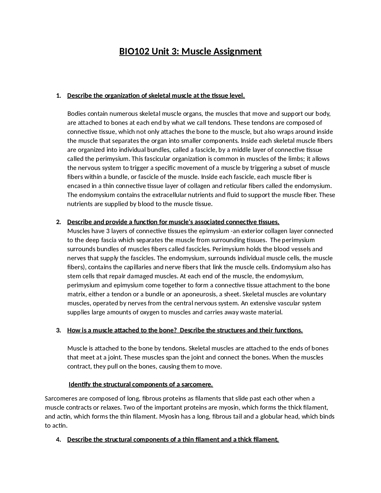
BIO102 Unit 3: Muscle Assignment (A GUARANTEED) <100% CORRECT> GRADED A+ | LATEST SOLUTIONS | 1. Describe the organization of skeletal muscle at the tissue level. Bodies contain numerous skeletal m... uscle organs, the muscles that move and support our body, are attached to bones at each end by what we call tendons. These tendons are composed of connective tissue, which not only attaches the bone to the muscle, but also wraps around inside the muscle that separates the organ into smaller components. Inside each skeletal muscle fibers are organized into individual bundles, called a fascicle, by a middle layer of connective tissue called the perimysium. This fascicular organization is common in muscles of the limbs; it allows the nervous system to trigger a specific movement of a muscle by triggering a subset of muscle fibers within a bundle, or fascicle of the muscle. Inside each fascicle, each muscle fiber is encased in a thin connective tissue layer of collagen and reticular fibers called the endomysium. The endomysium contains the extracellular nutrients and fluid to support the muscle fiber. These nutrients are supplied by blood to the muscle tissue. 2. Describe and provide a function for muscle's associated connective tissues. Muscles have 3 layers of connective tissues the epimysium -an exterior collagen layer connected to the deep fascia which separates the muscle from surrounding tissues. The perimysium surrounds bundles of muscles fibers called fascicles. Perimysium holds the blood vessels and nerves that supply the fascicles. The endomysium, surrounds individual muscle cells, the muscle fibers), contains the capillaries and nerve fibers that link the muscle cells. Endomysium also has stem cells that repair damaged muscles. At each end of the muscle, the endomysium, perimysium and epimysium come together to form a connective tissue attachment to the bone matrix, either a tendon or a bundle or an aponeurosis, a sheet. Skeletal muscles are voluntary muscles, operated by nerves from the central nervous system. An extensive vascular system supplies large amounts of oxygen to muscles and carries away waste material. 3. How is a muscle attached to the bone? Describe the structures and their functions. Muscle is attached to the bone by tendons. Skeletal muscles are attached to the ends of bones that meet at a joint. These muscles span the joint and connect the bones. When the muscles contract, they pull on the bones, causing them to move. Identify the structural components of a sarcomere. Sarcomeres are composed of long, fibrous proteins as filaments that slide past each other when a muscle contracts or relaxes. Two of the important proteins are myosin, which forms the thick filament, and actin, which forms the thin filament. Myosin has a long, fibrous tail and a globular head, which binds to actin. 4. Describe the structural components of a thin filament and a thick filament. Each thick filament is approximately 15 nm in diameter, and each is made of several hundred molecules of myosin. Thin filaments, 7 nm in diameter, consist primarily of the protein actin, specifically fibrous actin. Each actin strand is composed of a string of subunits called globular actin. 5. What role does Calcium play in the activation of muscle contraction? When calcium binds to troponin, the troponin changes shape, removing tropomyosin from the binding sites. The sarcoplasmic reticulum stores calcium ions, which it releases when a muscle cell is stimulated; the calcium ions then enable the cross-bridge muscle contraction cycle. [Show More]
Last updated: 1 year ago
Preview 1 out of 10 pages
Reviews( 0 )
Document information
Connected school, study & course
About the document
Uploaded On
Mar 20, 2022
Number of pages
10
Written in

Additional information
This document has been written for:
Uploaded
Mar 20, 2022
Downloads
0
Views
55


 UHC Ethics & Compliance 2022.png)

.png)