Biology > STUDY GUIDE > BIOLOGY 235 MIDTERM 2 REVIEW STUDY GUIDE DOWNLOAD TO SCORE A (All)
BIOLOGY 235 MIDTERM 2 REVIEW STUDY GUIDE DOWNLOAD TO SCORE A
Document Content and Description Below
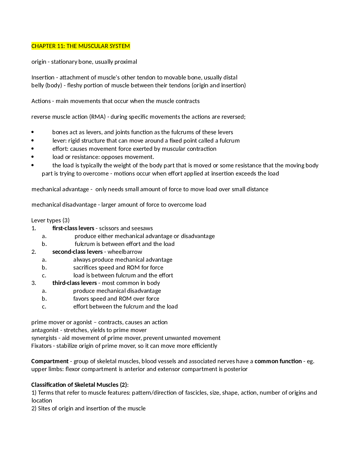
CHAPTER 11: THE MUSCULAR SYSTEM origin - stationary bone, usually proximal Insertion - attachment of muscle's other tendon to movable bone, usually distal belly (body) - fleshy portion of muscle be... tween their tendons (origin and insertion) Actions - main movements that occur when the muscle contracts reverse muscle action (RMA) - during specific movements the actions are reversed; bones act as levers, and joints function as the fulcrums of these levers lever: rigid structure that can move around a fixed point called a fulcrum effort: causes movement force exerted by muscular contraction load or resistance: opposes movement. the load is typically the weight of the body part that is moved or some resistance that the moving body part is trying to overcome - motions occur when effort applied at insertion exceeds the load mechanical advantage - only needs small amount of force to move load over small distance mechanical disadvantage - larger amount of force to overcome load Lever types (3) 1. first-class levers - scissors and seesaws a. produce either mechanical advantage or disadvantage b. fulcrum is between effort and the load 2. second-class levers - wheelbarrow a. always produce mechanical advantage b. sacrifices speed and ROM for force c. load is between fulcrum and the effort 3. third-class levers - most common in body a. produce mechanical disadvantage b. favors speed and ROM over force c. effort between the fulcrum and the load prime mover or agonist – contracts, causes an action antagonist - stretches, yields to prime mover synergists - aid movement of prime mover, prevent unwanted movement Fixators - stabilize origin of prime mover, so it can move more efficiently Compartment - group of skeletal muscles, blood vessels and associated nerves have a common function - eg. upper limbs: flexor compartment is anterior and extensor compartment is posterior Classification of Skeletal Muscles (2): 1) Terms that refer to muscle features: pattern/direction of fascicles, size, shape, action, number of origins and location 2) Sites of origin and insertion of the muscle Characteristics Used to Name Muscles: Direction Size Shape Action Number of origins Location Origin and insertion Muscles of the Head That Produce Facial Expressions ability to express emotion lie in subcutaneous layer Orifices: openings of the head - eyes, nose and mouth Sphincers: close orifices Dilators: dilate or open orifices orbicularis oculi - closes the eye occipitofrontalis - 2 parts: Frontal belly (frontalis) - superficial to frontal bone, draws scalp anteriorly + raises eyebrows, wrinkles skin of forehead horizontally Occipital belly (occipitalis) - posterior part, superficial to occiptal bone + draws scalp posteriorly held together by strong aponeurosis - covers superior and lateral surfaces of the skull buccinator - forms major muscular portion of the cheek Orbicularis oris - closes and protrudes lips, compresses lips against teeth and shapes lips during speech Zygomaticus major - draws angle of mouth superiorly and laterally (smiling) Muscles That Move the Mandible and Assist in Mastication Masseter - strongest muscle of mastication Origin: Maxilla and zygomatic arch Insertion: angle and ramus of mandible Action: Elevates mandible (closes mouth) temporalis Origin: temporal bone Insertion: coronoid process and ramus of mandible Action: elevates and retracts mandible Muscles of the Neck That Move the Head Balance and movement of the head on the vertebral column Sternocleidomastoid - two bellies: sternal head and clavicular head Origin: o sternal head --> manubrium of sternum o clavicular head --> medial third of clavicle Insertion: Mastoid process of temporal bone and lateral half of superior nuchal line of occipital bone Action: RMA: elevate sternum during forced inhalation o Bilaterally - flexes cervical portion of the vertebral column and flexes the head o Unilaterally - laterally flexes and rotates the head Muscles of the Abdomen That Protect Abdominal Viscera Anterolateral abdominal wall - composed of skin fascia and 4 pairs of muscles external oblique - superficial muscle - fascicles extend inferiorly and medially internal oblique - intermediate flat muscle - fascicles extend at right angles to external obliques rectus abdominis - long muscle extending whole length of anterior abdominal wall Origin: pubic crest and pubic symphysis Insertion: cartilages of ribs 5-7 and the xiphoid process Action: Flexes vertebral column (esp. lumbar), compresses abdomen to aid in defecation, urination, forced exhalation and childbirth (RMA: flexes pelvis on the vertebral column) transversus abdominis - deep muscle, fascicles directed transversely around abdominal wall Together with internal and external oblique, structural arrangement of muscle fascicles in different directions provide consideration protection to abdominal viscera Form linea alba During forceful exhalation – compresses abdomen Muscles of the Thorax That Assist in Breathing alter the size of thoracic cavity so breathing can occur Inhalation: breathing in (thoracic cavity increases in size) Exhalation: breathing out (thoracic cavity decreases in size) diaphragm - important muscle that powers breathing - separates thoracic and abdominal cavities Convex superior surface - forms floor of thoracic cavity Concave inferior surface - forms roof of abdominal cavity Contraction of diaphragm causes it to flatten and increases vertical height in thoracic cavity (inhalation) Relaxation of diaphragm causes it to move superiorly and decrease in height in thoracic cavity (exhalation) Intercostals - involved in breathing - in intercostal spaces (spaces between ribs) 1. external intercostals - 11 pairs occupy superficial layer, fibers run in an oblique direction interiorly and anteriorly from rib above and rib below a. Elevate ribs during inhalation - expends thoracic cavity b. During relaxation: depress ribs and decreases anteroposterior and lateral dimensions of thoracic cavity (exhalation) 2. internal intercostals - 11 pairs occupy intermediate layer of intercostal spaces, fibers run at right angles to the external intercostals a. Draw adjacent ribs together during forced exhalation - helps to decrease size of thoracic cavity Muscles of the Thorax That Move the Pectoral Girdle scapular movements accompany humeral movements to i [Show More]
Last updated: 1 year ago
Preview 1 out of 78 pages
Reviews( 0 )
Document information
Connected school, study & course
About the document
Uploaded On
Jan 08, 2022
Number of pages
78
Written in
Additional information
This document has been written for:
Uploaded
Jan 08, 2022
Downloads
0
Views
67

