Clinical Chemistry > EXAM > VET 1335 Clinical Pathology I Midterm Over 150 Questions and Answers (All)
VET 1335 Clinical Pathology I Midterm Over 150 Questions and Answers
Document Content and Description Below
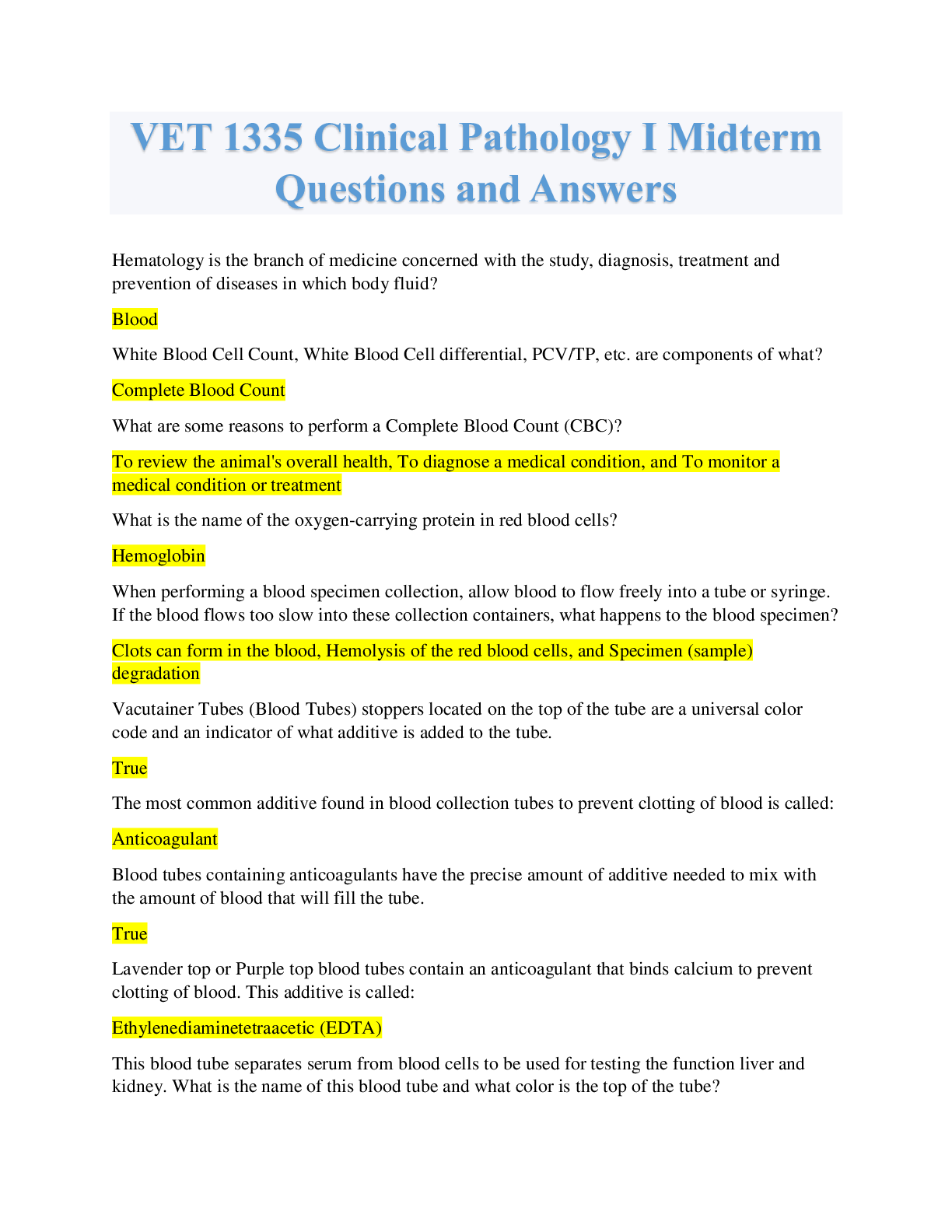
SAMPLE QUESTIONS HERE: VET 1335 Clinical Pathology I Midterm Questions and Answers Hematology is the branch of medicine concerned with the study, diagnosis, treatment and prevention of diseases in ... which body fluid? White Blood Cell Count, White Blood Cell differential, PCV/TP, etc. are components of what? What are some reasons to perform a Complete Blood Count (CBC)? What is the name of the oxygen-carrying protein in red blood cells? When performing a blood specimen collection, allow blood to flow freely into a tube or syringe. If the blood flows too slow into these collection containers, what happens to the blood specimen? Vacutainer Tubes (Blood Tubes) stoppers located on the top of the tube are a universal color code and an indicator of what additive is added to the tube. The most common additive found in blood collection tubes to prevent clotting of blood is called: Blood tubes containing anticoagulants have the precise amount of additive needed to mix with the amount of blood that will fill the tube. Lavender top or Purple top blood tubes contain an anticoagulant that binds calcium to prevent clotting of blood. This additive is called: This blood tube separates serum from blood cells to be used for testing the function liver and kidney. What is the name of this blood tube and what color is the top of the tube? Components of blood are: Which component of blood fights infection and disease? This blood component is the extracellular matrix of blood cells. It is mostly water and contains dissolved proteins. This is the rupture of red blood cells with the release of hemoglobin and the intracellular components into the plasma. Appears pale red to cherry red in color. What is this condition called? This is the presence of high levels of bilirubin (old RBCs that are broken down by the liver). The serum or plasma will be the color of bright yellow. What is this condition called? This is the presence of excess lipids or fats in the blood. This condition causes the plasma or serum to appear white or milky. In some breeds of dogs this condition is normal. What is this condition called? This is the production of blood cells and platelets in the red bone marrow. What is the term for this production? All blood cells arise from Pluripotent hematopoietic stem cells. Part of Hematopoiesis, the white blood cells (Leukocytes) is split into two characteristic groups. This group of two cells contains some granules in the cell and have a mononuclear (one solid) nucleus. The two cells in this group are the Monocytes and Lymphocytes. What characteristic group do these cells belong to? These white blood cells have many granules and their nucleus contain multiple lobes (polymorphonuclear). The cells names are neutrophils, eosinophils, and basophils. What characteristic group do these cells belong to? This WBC is an agranulocyte, has a mononuclear nucleus that stains purple and the most important cell of the immune system! What is the name of the WBC? This WBC is a granulocyte and is the most numerous WBC. The cell's job is to phagocytize and destroy bacteria. The nucleus is polymorphonuclear. What is the name of the WBC? This WBC is an agranulocyte and is the largest of the leukocytes. A unique feature is the nucleus sometimes is kidney shaped, and it transforms into macrophage as it gets older and phagocytizes cells. What is the name of the WBC? This WBC is a granulocyte and is composes 1-4% of all WBCs (leukocytes). The cell ends allergic reactions and is seen in parasitic infections. The granules will stain, pinkish-red and each species of animals have their own unique cell. What is the name of the WBC? This WBC is a granulocyte and is rarely seen in blood smears, but the nucleus usually has two lobes. The granules secreted histamines and seen in inflammations. What is the name of this WBC? During Erythropoiesis, Erythrocytes (RBCs) are called Reticulocytes when first formed in the bone marrow and mature into RBCs. This process of Reticulocytes maturing into RBCs takes how many days in the bloodstream? What organ in the body plays a role in the development of RBC's (Erythropoiesis) by excreting a hormone called Erythropoietin? What is the term for the development of platelets (Thrombocytes)? In the Monocyte, what characteristic cell structure is commonly noted in the cytoplasm of the cell? Part of a Complete Blood Count (CBC) includes an examination of a blood smear that is usually performed by a qualified veterinary technician. Blood smears should be made and stained how long after the sample is collected? If blood smears are not made and stained immediately, what is the name of the additive in the lavender blood tube that the WBCs and other cells will degenerate rapidly in? Other names for a blood smear is blood film or wedge smear. Blood smears are checked for types of WBCs, shapes of RBCs, and the presence of platelets. This procedure is called. The structure and shape of all cells (WBCs, RBCs, Platelets) is termed: Creating a blood smear is by placing a drop of blood on one end of a glass slide, and using a spreader/push slide to disperse the blood over the slide's length. What is the goal of this procedure regarding the cells in the blood? The single layer of cells (monolayer), are spaced far enough apart to be counted and differentiated on the slide under a microscope. On what part of the slide is the monolayer of cells found? How many blood smears are prepared for a patient? When drying a blood smear, the blood on the slide has to be dried quickly instead of slowly. Why? What are some characteristics of a well-made slide? Is dirt or grease on the slide, a drop of blood too small, a chipped or rough edge of spreader/push slide indications for sources of error that could cause inaccurate results? Stains used in the staining of blood smears will stain the different structures of the cell. This is to make the cell structures more visible under microscopic exam. There are three steps to the staining process of a blood smear called Wright's Stain Manual Method. The first step of this process is to preserve the morphology of the blood cells. What is the name of the first reagent in the staining the blood smear? This reagent is a dye that produces colors base on the ionic charges of the stain and various components of cells. This reagent has negative charges and stains the cell components and orange to pink color. What is the name of this reagent in the second step of the staining process? The last reagent in the staining process of a blood smear slide contains ions that are positively charged and will stain the acid cell components varying shades of blue. What is the name of this reagent? In the three steps of staining, holding the edge of slide and dip: Once the blood smear slide has been stained and dried, it is placed under the microscope on low power (10X). The whole slide is scanned for what first? Performing the WBC differential count is performed: Each WBC type is recorded as a percentage and this procedure is called a Relative WBC count. Monocytes transform into macrophages and become eating machines. Unique characteristics of monocytes are: Monocytes after receiving a chemical message from chemokines, the cell does not migrate out of the blood vessels into the damaged tissue. Monocytopenia can be seen in acute infections or inflammation and stress. What does the term Monocytopenia mean? Increased numbers of Monocytes are seen in the blood where there are chronic inflammatory processes taking place. This increased in the number of Monocytes in the blood is called: The most important WBC in the immune system is the: Lymphocyte What are the two main classes of lymphocytes? A unique feature of a Lymphocyte is the large round nuclei and a small rim of blue cytoplasm. This Lymphocyte is commonly seen in a young animals that has been recently vaccinated due to antigen stimulation. The characteristics are a large cell, coarse chromatin and have a deep blue cytoplasm. What is the name of this unique Lymphocyte? Reactive lymphocyte When blood work is characterized by neutrophilia, monocytosis, eosinopenia and lymphopenia all associated with stress from a disease, this is called a: Stress Leukogram All white blood cells are produced and derived from cells in the bone marrow is known as: Hematopoietic This laboratory procedure determines the percentage of each type of white blood present in the blood. WBC differential This laboratory procedure measure the number of white blood cells in a volume of blood. WBC count The principle of the Lecuko-TIC procedure is counting of WBC nuclei. What is the name of the piece of equipment used in counting of WBC nuclei? Hemacytometer The Leuko-Tic principle is microscopic counting of WBCs after the lysis of RBCs and the fixing of WBC nuclei. Is the hemacytometer disposed of in the biohazard container or waste paper basket after the procedure? No EDTA fresh blood is required when performing the Leuko-TIC procedure. If a chamber is over charged, this is considered a source of error. As with any diagnostic procedure there has to be quality control. With WBC counts, the number of WBC nuclei in each of 8 WBC squares on the hemacytometer chambers should agree within how many WBC nuclei (cells)? 10 Another quality control with WBC counts is the hemacytometer chamber #1 should agree within _______ WBC nuclei (cells) of chamber #2. 15 Leukocytosis is an increase of WBC count above a species normal reference range. What are some clinical findings that could cause this condition? Acute inflammatory diseases and Bacterial infections Left shift is an increase in the number of immature leukocytes in the peripheral blood, particularly neutrophil band cells. If a WBC differential count shows band neutrophil count higher than segmented neutrophils this usually indicates severe inflammation. There is morphologic changes too and are termed "Toxic" to the WBCs. What is the name of this left shift condition? Degenerative Left shift Nucleated Red Blood cells (nRBCs) are immature red blood cells with a nucleus present. During the maturation process the nucleus is expelled before the RBC is released in blood circulation. The maturation process of RBCs is called what? Erythropiesis Low numbers of nRBCs are naturally seen in cats What diseases processes cause the bone marrow to produce RBCs and immature RBCs being released into circulation before they are mature? Anemia, Cancers involving bone marrow and Myelofibrosis What WBC does nRBCs must be distinguished from? Lymphocyte What are some characteristics of nRBCs? Nuclear chromatin is very dense and smooth and Cytoplasm can be dark blue or purple WBC counts are actually of WBC and nRBC nuclei counted together. nRBCs are not lysed and are distinguishable from WBC nuclei. The WBC count must be corrected for the number of nRBC in the blood circulation. What instrument is used for WBC counts? Hemacytometer What species of animal have naturally low numbers of nRBCs? Camelid WBC count corrections are not performed when nRBCs are less than 5 nRBCs per 100 WBCs. The Packed Cell Volume (PCV) measures the percentage of ______________ to the total blood volume. Red blood cells (RBCs) Red blood cells (RBCs) carry what throughout the body? Oxygen The purpose of a Packed Cell Volume (PCV) is to: Measure the volume of red blood cell component of blood and Used as an indicator of hemoglobin What kind of tube is a Packed Cell Volume (PCV) measured in? Microhematocrit tube Packed Cell Volume (PCV) is determined by measuring the ________ of the red blood cell column in a microhematocrit tube. Height The space occupied by the red blood cells in the microhematocrit tube is measured in what unit? mm How many microhematocrit tubes are drawn up when preparing for a Packed Cell Volume (PCV) procedure? 2 Blood with the additive EDTA is required to run a PCV. Once the hematocrit tube has been spun down in the centrifuge, there are three layers seen. What is the name of the three layers? Packed Red cells, Plasma and Buffy coat Packed cell volume (PCV) ranges in animals are all the same. Normal values for PCV may differ from laboratory to laboratory. When reading a PCV, a lower than normal reference range for any species of animal may indicate the following: Vitamin or mineral deficiencies, Long-term blood loss and Anemia (insufficient supply of healthy RBCs) When reading a PCV, a higher than normal reference range for any species of animal may indicate the following: An increasing amount of EDTA (anti-coagulant) in the blood will decrease the PCV reading as a result of: Sealing the end of the capillary tube is important, so there is no loss of blood from the tube during centrifugation. Plasma is mostly composed of what? The plasma portion of a hematocrit tube is measured by an instrument called: When reading the plasma portion of a hematocrit tube, it is called reading the: To read a Total Protein from a hematocrit tube, the tube needs to broken above what layer in the tube? In the PCV layers of cells and plasma, which layer contains WBCs and Platelets? Plasma layer of the PCV can indicate a disease process by the color of the plasma. When measuring a TP with the refractometer it is important to obtain a drop of plasma from the broken end of the tube. When reading a TP with the refractometer you need a weak source of light. What is the value or unit that is recorded when reading a TP off a refractometer? What are some clinical findings from a decreased value of a TP? Increased values of a TP could indicate dehydration. A refractometer must be regularly calibrated Neutrophils, Eosinophils, Basophils contain many granules and fall into the class of WBCs also known as Leukocytes called: Lymphocytes and Monocytes contain few granules and fall into the class of WBCs also known as Leukocytes called: When looking at WBCs under a microscope and identifying the different kinds of WBCs according to their characteristics (size, shape, number of granules, chromatin pattern, etc.), this is called a: Retics must be stained with new methylene blue in order to differentiate between aggregate and punctate retics. When neutrophils (a WBC) is young in the bloodstream, the cell is called a "Band" cell. Due to there is only one nucleus usually in a horseshoe shape. As the neutrophil goes through the "maturation" or process of maturing, the nucleus of the cell starts segmenting into 2-6 lobes. What is the cell called at this stage of maturation? Neutrophils are drawn to the site of inflammation through chemical messaging. Once neutrophils reach the site of inflammation, the cell will start to digest the bacteria. This process is known as: There is species variation in most WBCs. The canine neutrophil has a unique appendage feature on the nucleus indicating that the canine is female. What is the name of this unique characteristic? Increased or decreased WBCs in the blood is a clinical indication of a disease process. An increased in numbers of neutrophils is called: A decrease in WBCs is called Some causes of Neutrophilia are: Feline Leukemia virus can cause Neutropenia. After an acute infection, it takes how many hours for the bone marrow to increase the number of neutrophils in the blood? Eosinophils combat multicellular parasites and certain infections. The WBC also is associated with control mechanisms for allergies and asthma. In eosinophils, there is a marked species variation by the number, size, and shapes of granules. Canine eosinophils have the most interspecies variation in the granule size, number, and shape. In the canine eosinophils, what cell characteristic is very commonly found? The granules found in Feline eosinophils are: The granules in the Equine are very large and look like a: A dog has blood work performed, and the results show a high count of eosinophils. This high number of eosinophils is called: Acute stress reactions, acute inflammation, Cushing syndrome with steroid therapy can cause a decrease of eosinophils circulating in the blood. This decrease of eosinophils is called: In healthy animals, basophils are rarely seen in a WBC differential. Basophils granules contain histamine and heparin. The histamine plays a role in acute allergy, and the heparin inhibits coagulation. The WBC Monocytes unique characteristics are: What size end-to-end volume capillary tube is used for the Leuko-TIC procedure? Once placing the end-to-end volume capillary tube in the vial, the vial is shook to flush all the blood from the vial. Then we wait for 30 seconds. What are we waiting for? Once the liquid in the vial has been pulled up into a capillary tube, the liquid is then loaded into the counting chambers. What is this procedure called? If a chamber is over charged, this is considered a source of error. When beginning counting the WBC nuclei in the Hemacytometer chambers, what objective should the microscope be on? It is recommended to reduce the condenser and dimmer on the microscope when beginning a WBC count. What is the name of device used in counting in WBC Differentials and Counts? When beginning counting the WBC nuclei (cells) in the chambers of the hemacytometer, where do you begin counting? The hemacytomer chamber has four corner squares. Each square contains how many small squares for counting the WBC nuclei (cells)? Count WBC nuclei (cells) touching the border lines on just ______ sides of each square. You repeat the WBC nuclei count procedure in all 4 squares of the hemacytometer chamber. As with any diagnostic procedure there has to be quality control. With WBC counts, the number of WBC nuclei in each of 8 WBC squares on the hemacytometer chambers should agree within how many WBC nuclei (cells)? Another quality control with WBC counts is the hemacytometer chamber #1 should agree within _______ WBC nuclei (cells) of chamber #2. Uneven distribution of the quality control numbers in a WBC count is typically from dirty hemacytometer or coverslip. There are many sources of error involving a WBC count preparation. Which of the following are sources of error? If the WBC count is decrease or below a species reference norms for WBC count, this is called: Leukocytosis is an increase of WBC count above a species normal reference range. What are some clinical findings that could cause this condition? Left shift is an increase in the number of immature leukocytes in the peripheral blood, particularly neutrophil band cells. If a WBC differential count shows band neutrophil count higher than segmented neutrophils this usually indicates severe inflammation. There is morphologic changes too and are termed "Toxic" to the WBCs. What is the name of this left shift Reticulocytes ( polychromatophils) are: In the process of red blood cell formation in the bone marrow (erythropoiesis) is where the reticulocytes develop. The nucleus is already ejected when the red blood cell matures to the reticulocyte stage. What are polychromatophils? A polychromatophil appears to be bluer and tends to be larger than a normal red blood cell. When the polychromatophilic are stained with New Methylene Blue stain, the stain highlights the ribosomes in the cytoplasm. When this occurs the polychromatophils are then called: Whenever there is anemia present, a retic count is indicated to evaluate the bone marrow response and aid in classifying the type anemia. Retics are classified into two types of immature anucleated RBCs. This classification is dependent on how much RNA is left and how it stains. Aggregate retics are more immature and contain more RNA. The punctate retics have less RNA which makes the cell more mature. When aggregate retic are stained in New methylene blue, they look like Polychromatophils that was a stain in Wrights stain. Punctate retics stain red in a Wright stain smear because they have to little _________ to stain purple. Back to retics themselves. Retics are really non-nucleated red blood cells that contain remnants of RNA material that is called: Bone marrow or regenerative response to anemia is assessed by identifying reticulocytes in the peripheral blood. Normally healthy dog and cats have low numbers of retics. A reticulocyte count is the most effective measure of: An elevated count of retics in response to anemic stress is called: What other immature types of cells may be seen with retics as the bone marrow races to deliver cells at a rapid rate? Species of animals differ in their regenerative response in both degrees and the types of immature RBCs the bone marrow releases. Increase reticulocyte counts may be referred to as: Regenerative anemia where the retic count is increased can be caused by what states: In the bone marrow regenerative response to anemia, there are other remanent RNA or ribosomes features inside the RBC when it is quickly released from the bone marrow before it is fully mature. We know about the nucleated RBCs, but there are two other features. What are these features or inclusions? A decrease in reticulocyte counts is referred to as: Non-Regenerative anemia means that the bone marrow is not responding to the blood loss or anemia. Immature RBCs that contain large amounts of RNA are called: In aggregate retics, what type of stain is required to stain the RNA blue? The large amount of RNA in aggregate retics will offset the red of the hemoglobin. Thus, leaving a purple color of the cytoplasm. The change in the RBC color is called: In cats, aggregate reticulocytes mature into punctate retics within: Because aggregate retics mature so quickly into punctate retics in the cat, only aggregate retics are counted. With the dog, both aggregate and punctate retics are counted during a retic count. Which species are retic counts not performed because there are too few retics released in response to anemia? In the ruminants, no reticulocytes or polychromatophils circulate in the blood, so retics are not counted. Which species of animals respond poorly to blood loss anemia which there is a poor regenerative response, and it can take weeks to a month for this species to recover from blood loss. To perform a retic count, what type of well-mixed blood is required? In preparing for a retic count, how many drops of blood are placed into a clean test tube by a pipette? Once the New Methylene blue stain and blood are gently mixed in the test tube, how long do let the mixture incubate at room temperature? Once the incubation process is complete, then the test tube is shaken to mixed the blood and stain again. Are the slides for retic count prepared similarly to a differential smear slide? When making the slide smears for the retic count, the slide needs to be made thick. It is acceptable to dry the slide using a hair dryer which is much faster than air drying. Once the slide is prepared and dried for the retic count, it is placed under what objective on the microscope? When viewing the slide under the microscope, the circular viewing area is split up into how many sections visually? The retic count is starting in the circular viewing area left to right. The WBC differential counter can be used to perform a retic count. When counting for retics, all RBCs are counted including the retics until how many cells are reached? When performing a retic count, both prepared slides are counted Once the retic counting is completed, the two slides should agree within how many ___ %? What are some sources of error when performing a retic count? Like with all hematology cell counts, there has to be an allowance for corrections. The retic count is only corrected when a patient PCV is: Absolute Retic counts provide a more accurate method of whether or not anemia is regenerative and the adequacy of the regenerative response. It is useful to have a baseline hematocrit (PCV) from a patient presented with an illness, This baseline is used when diagnosing what type of anemia the patient has and if it is regenerative or non-regenerative. [Show More]
Last updated: 1 year ago
Preview 1 out of 19 pages
Reviews( 0 )
Document information
Connected school, study & course
About the document
Uploaded On
Mar 11, 2022
Number of pages
19
Written in

Additional information
This document has been written for:
Uploaded
Mar 11, 2022
Downloads
0
Views
84


.png)